
















Copyright: ©Author(s) 2026.
World J Stem Cells. May 26, 2026; 18(5): 117584
Published online May 26, 2026. doi: 10.4252/wjsc.v18.i5.117584
Published online May 26, 2026. doi: 10.4252/wjsc.v18.i5.117584
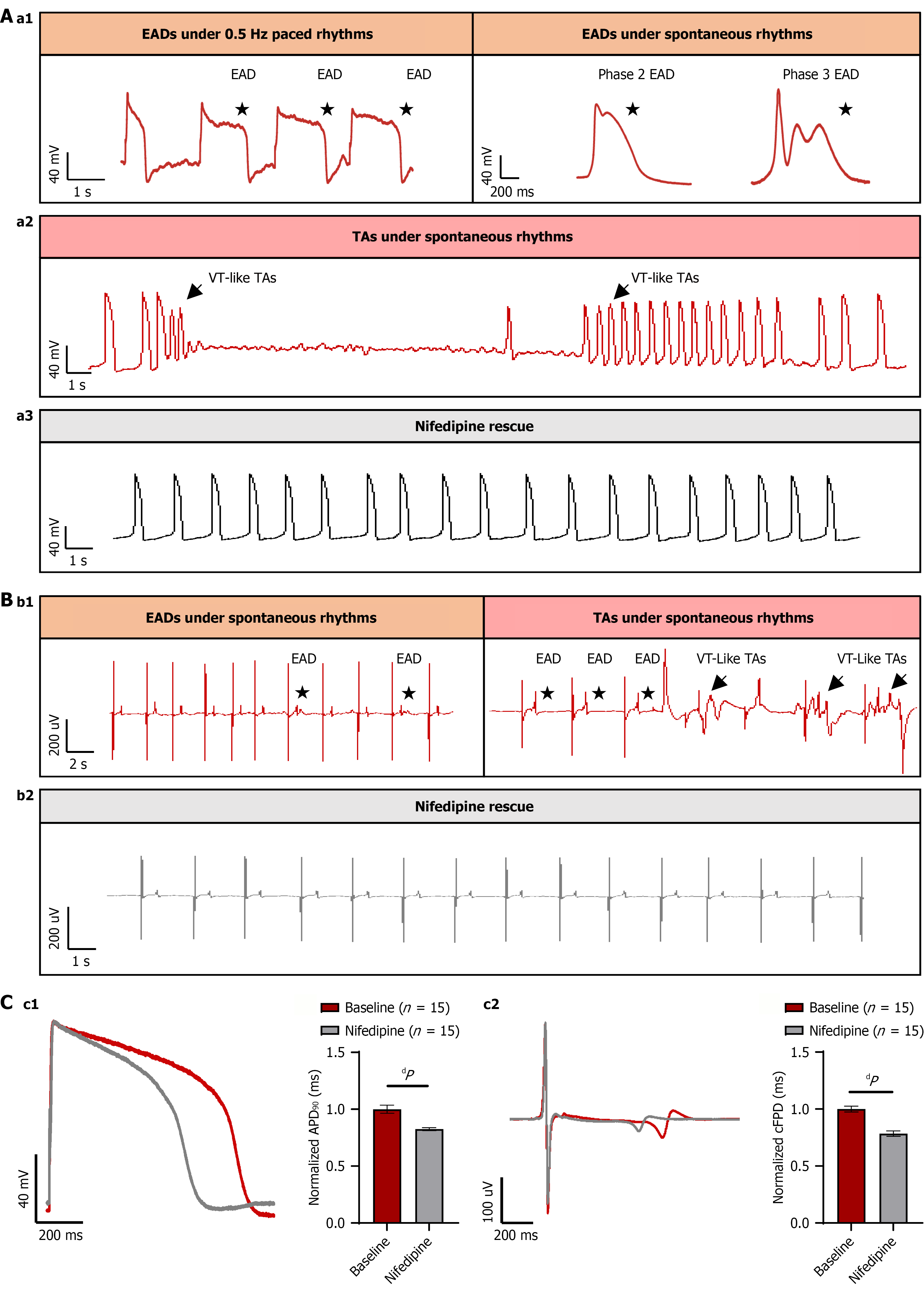
Figure 5 Arrhythmogenic behavior of family trio induced pluripotent stem cell-derived cardiomyocytes.
A: Representative early after
- Citation: Li Q, Guo CT, Ren JC, Wang YF, Zhao ZJ, Lv CH, Wang QY, Liu Q, Li K, Yang J, He R, Liu FL, Lv TT, Zhang P. Calcium dysregulation underlies phenotypic diversity in LQT2: Insights from induced pluripotent stem cell-derived cardiomyocytes of a KCNH2 p.Y427H family trio. World J Stem Cells 2026; 18(5): 117584
- URL: https://www.wjgnet.com/1948-0210/full/v18/i5/117584.htm
- DOI: https://dx.doi.org/10.4252/wjsc.v18.i5.117584