
















Copyright: ©Author(s) 2026.
World J Stem Cells. May 26, 2026; 18(5): 117584
Published online May 26, 2026. doi: 10.4252/wjsc.v18.i5.117584
Published online May 26, 2026. doi: 10.4252/wjsc.v18.i5.117584
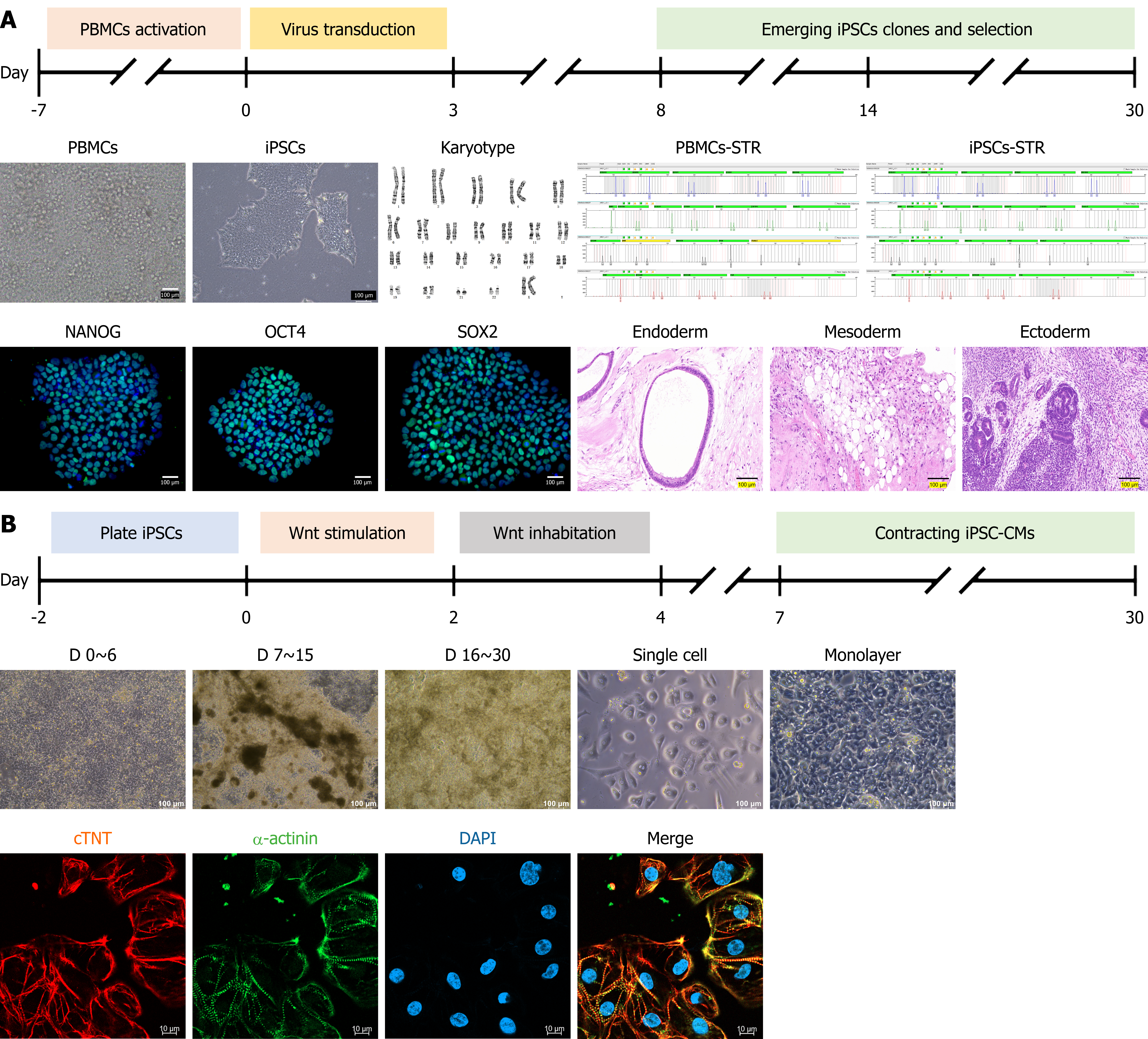
Figure 2 Generation and characterization of family trio induced pluripotent stem cell-derived cardiomyocytes.
A: Reprogramming process: Representative bright-field images of peripheral blood mononuclear cells and 10th-passage induced pluripotent stem cells (iPSCs). G-banding karyotyping and short tandem repeat profiling confirm genomic stability. Immunofluorescence staining shows robust expression of pluripotency markers (NANOG, OCT4, SOX2) in green, with a DAPI counterstain in blue. Teratoma formation experiments in immunodeficient mice demonstrate iPSCs differentiation into all three germ layers: Endoderm (respiratory epithelium), mesoderm (adipocytes), and ectoderm (neuronal cells), confirming pluripotency. Scale bars: 100 μm; B: Differentiation process: Mor
- Citation: Li Q, Guo CT, Ren JC, Wang YF, Zhao ZJ, Lv CH, Wang QY, Liu Q, Li K, Yang J, He R, Liu FL, Lv TT, Zhang P. Calcium dysregulation underlies phenotypic diversity in LQT2: Insights from induced pluripotent stem cell-derived cardiomyocytes of a KCNH2 p.Y427H family trio. World J Stem Cells 2026; 18(5): 117584
- URL: https://www.wjgnet.com/1948-0210/full/v18/i5/117584.htm
- DOI: https://dx.doi.org/10.4252/wjsc.v18.i5.117584