
















Copyright: ©Author(s) 2026.
World J Stem Cells. May 26, 2026; 18(5): 117241
Published online May 26, 2026. doi: 10.4252/wjsc.v18.i5.117241
Published online May 26, 2026. doi: 10.4252/wjsc.v18.i5.117241
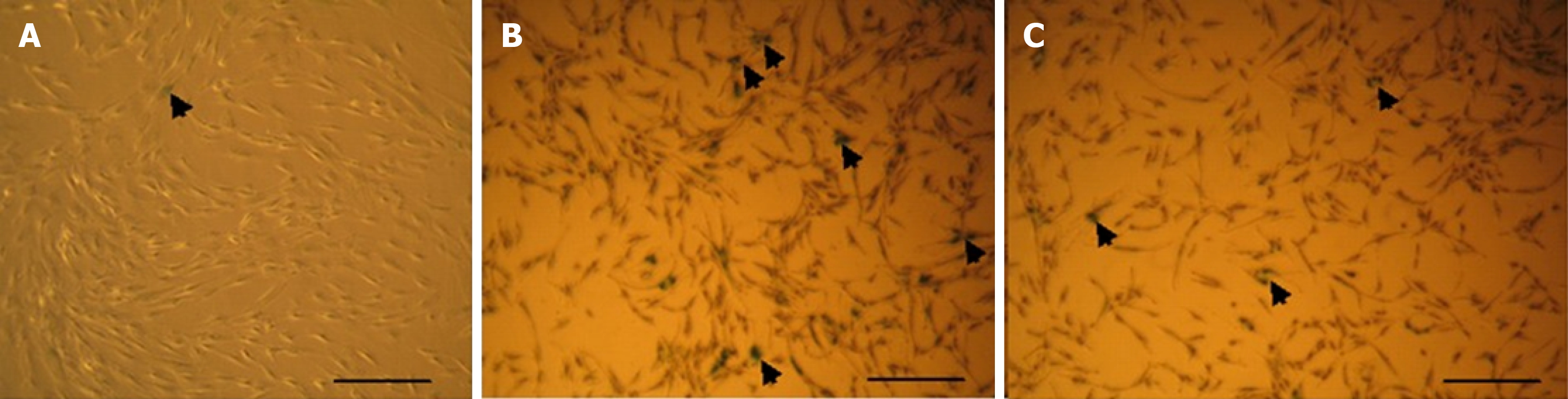
Figure 8 Representative micrographs of senescence-associated β-galactosidase staining.
The images were captured at 10 × magnification. Arrow head represents senescence cells. A: Young adipose tissue derived mesenchymal stem cells (ADMSCs) displayed few senescence-associated β-galactosidase (SA-β-gal)-positive cells, indicating a low baseline level of senescence; B: Aged normoxia ADMSCs exhibited a markedly higher proportion of SA-β-gal-positive cells; C: Aged hypoxia ADMSCs demonstrated a visibly reduced proportion of SA-β-gal-positive cells compared with their normoxic aged counterparts.
- Citation: Bashir B, Choudhery MS, Hussain I. Hypoxic preconditioning restores the regenerative potential of aged adipose tissue derived mesenchymal stem cells. World J Stem Cells 2026; 18(5): 117241
- URL: https://www.wjgnet.com/1948-0210/full/v18/i5/117241.htm
- DOI: https://dx.doi.org/10.4252/wjsc.v18.i5.117241