
















Copyright: ©Author(s) 2026.
World J Stem Cells. May 26, 2026; 18(5): 117241
Published online May 26, 2026. doi: 10.4252/wjsc.v18.i5.117241
Published online May 26, 2026. doi: 10.4252/wjsc.v18.i5.117241
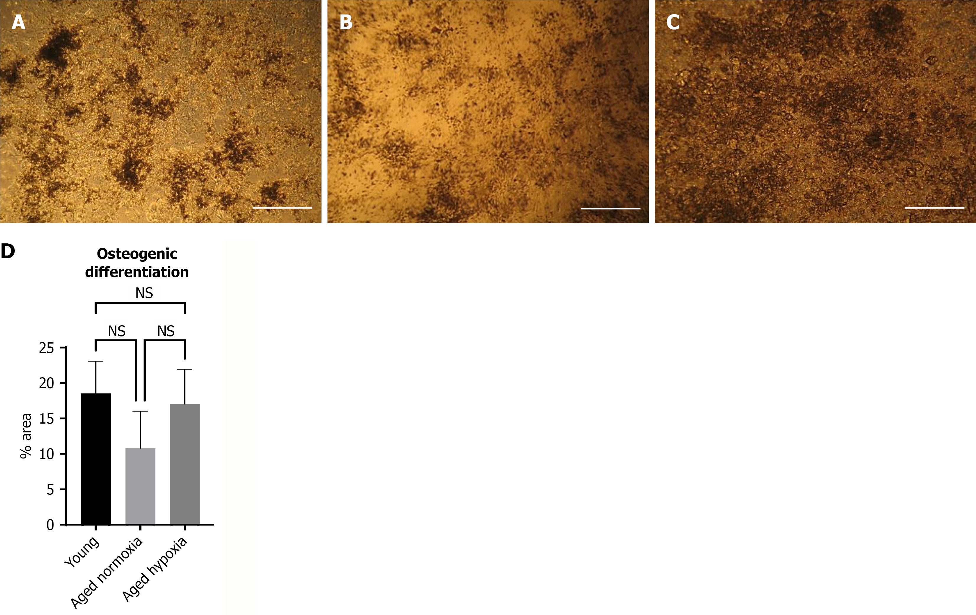
Figure 4 Osteogenic differentiation of adipose tissue derived mesenchymal stem cells assessed by von Kossa staining after 21 days of induction.
A: Young adipose tissue derived mesenchymal stem cells (ADMSCs) exhibited strong osteogenic differentiation with abundant mineralized nodules (20 ×); B: Aged normoxic ADMSCs showed markedly reduced mineralization (20 ×); C: Aged-hypoxic ADMSCs demonstrated mineralized nodules at levels comparable to young ADMSCs (20 ×); D: Quantification of osteogenic differentiation potential of young ADMSC, aged normoxia ADMSCs and aged hypoxia ADMSCs. Data are presented as mean ± SD (n = 3). NS: Not significant.
- Citation: Bashir B, Choudhery MS, Hussain I. Hypoxic preconditioning restores the regenerative potential of aged adipose tissue derived mesenchymal stem cells. World J Stem Cells 2026; 18(5): 117241
- URL: https://www.wjgnet.com/1948-0210/full/v18/i5/117241.htm
- DOI: https://dx.doi.org/10.4252/wjsc.v18.i5.117241