
















Copyright: ©Author(s) 2026.
World J Stem Cells. May 26, 2026; 18(5): 117045
Published online May 26, 2026. doi: 10.4252/wjsc.v18.i5.117045
Published online May 26, 2026. doi: 10.4252/wjsc.v18.i5.117045
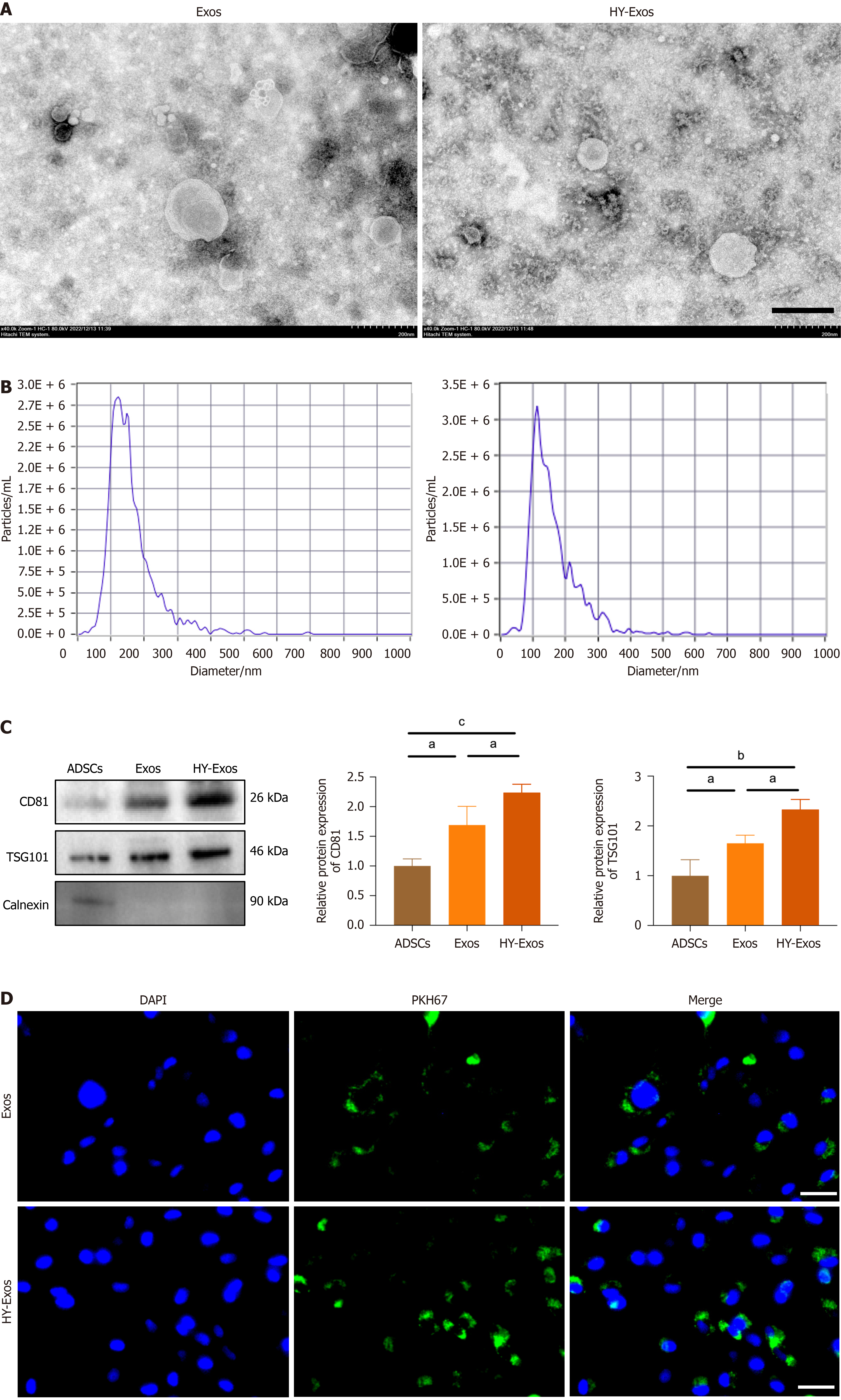
Figure 1 Hypoxia promotes exosomal release from adipose-derived stem cells and uptake by bone marrow mesenchymal stem cells.
A: Representative transmission electron microscopy images showing exosome morphology (scale bar: 200 nm); B: Nanoparticle tracking analysis showing size distribution of adipose-derived stem cells-derived exosomes; C: Western blot analysis and quantification of exosomal markers CD81 and tumor susceptibility gene 101, with calnexin as a negative control (n = 3, one-way ANOVA); D: Fluorescence microscopy images showing colocalization of exosomes with bone marrow mesenchymal stem cells. Nuclei are stained with DAPI (blue), PKH67-labeled exosomes are shown in green, and merged images are presented (scale bar: 100 μm). aP < 0.05; bP < 0.01; cP < 0.001. Exos: Exosomes; HY-Exos: Hypoxia-preconditioned exosomes; ADSCs: Adipose-derived stem cells; TSG101: Tumor susceptibility gene 101.
- Citation: Taxi W, Sun J, Zhu YH, Zhang TC, Guo Y, Pan YL, Liu MM, Chen X, Zhang CL, Tong K, Wang LN, Jiang T, Ma Y. Hypoxia-preconditioned adipose-derived stem cell exosomes promote osteogenic differentiation and bone formation in bone marrow mesenchymal stem cells through the Piezo1/Ca2+ signaling axis. World J Stem Cells 2026; 18(5): 117045
- URL: https://www.wjgnet.com/1948-0210/full/v18/i5/117045.htm
- DOI: https://dx.doi.org/10.4252/wjsc.v18.i5.117045