
















Copyright: ©Author(s) 2026.
World J Stem Cells. May 26, 2026; 18(5): 116611
Published online May 26, 2026. doi: 10.4252/wjsc.v18.i5.116611
Published online May 26, 2026. doi: 10.4252/wjsc.v18.i5.116611
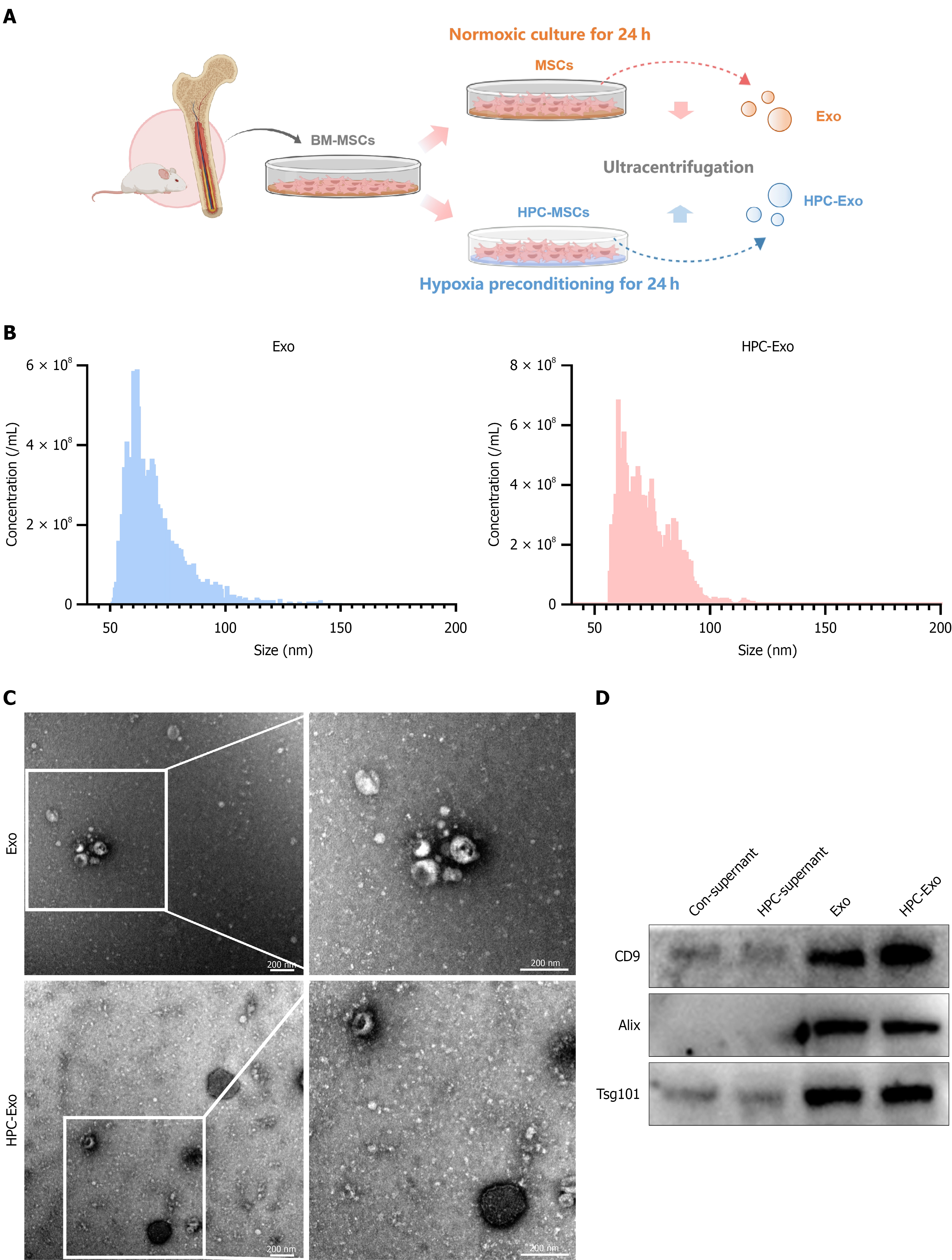
Figure 1 Characterization of exosome and hypoxia-preconditioned mesenchymal stem cell-derived exosomes.
A: Schematic diagram of the isolation process for exosomes derived from mesenchymal stem cells; B: Size distribution chart of exosome (Exo) and hypoxia-preconditioned mesenchymal stem cell-derived Exo; C: Transmission electron micrographs of Exo and hypoxia-preconditioned mesenchymal stem cell-derived Exo. Scale bar: 200 nm; D: Western blot analysis of Alix, Tsg101, and CD9 expression in Exo and HPC-Exo. BM-MSC: Bone marrow mesenchymal stem cell; Exo: Normoxic exosomes; HPC-Exo: Hypoxia-preconditioned mesenchymal stem cells-derived exosomes; MSCs: Mesenchymal stem cells.
- Citation: Wen W, Liu CX, Wang Y, Lu XJ, Yang C, Chen SJ, Jin ZT, Qu MY, Deng JY, Zhang Z. Hypoxia-preconditioned mesenchymal stem cell-derived exosomes attenuate myocardial ischemia/reperfusion injury by miR-29b-3p via PINK1/Parkin-mediated mitophagy. World J Stem Cells 2026; 18(5): 116611
- URL: https://www.wjgnet.com/1948-0210/full/v18/i5/116611.htm
- DOI: https://dx.doi.org/10.4252/wjsc.v18.i5.116611