
















Copyright: ©Author(s) 2026.
World J Stem Cells. May 26, 2026; 18(5): 116280
Published online May 26, 2026. doi: 10.4252/wjsc.v18.i5.116280
Published online May 26, 2026. doi: 10.4252/wjsc.v18.i5.116280
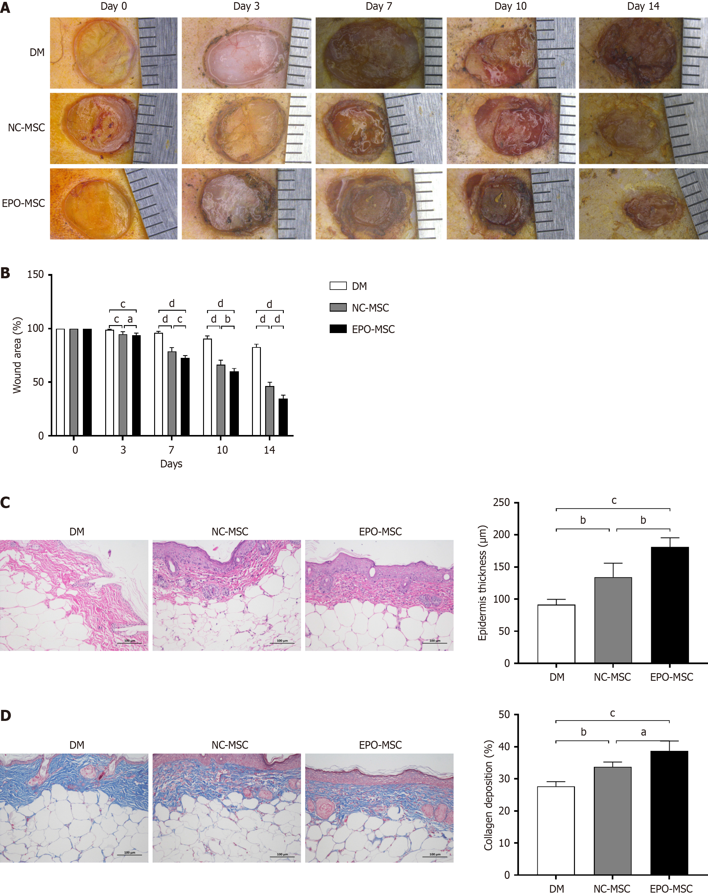
Figure 2 Effects of erythropoietin-overexpressing mesenchymal stem cells on skin wound healing in diabetic mice.
A: Representative macroscopic images of full-thickness dorsal skin wounds in streptozotocin-induced diabetic mice treated with vehicle (diabetes mellitus group), negative control mesenchymal stem cells, or erythropoietin-overexpressing mesenchymal stem cells at days 0, 3, 7, 10, and 14 after wounding; B: Quantification of wound area over time, expressed as a percentage of the initial wound area for the diabetes mellitus, negative control mesenchymal stem cell, and erythropoietin-overexpressing mesenchymal stem cell groups; C: Representative hematoxylin and eosin staining images of skin wound sections at day 14 and quantification of epidermal thickness; D: Representative Masson’s trichrome staining images of skin wound sections at day 14 and quantification of collagen deposition. Data are presented as mean ± SD. Superscript letters denote statistically significant differences between the indicated groups (aP < 0.05, bP < 0.01, cP < 0.001, dP < 0.0001; one-way ANOVA followed by Tukey’s post hoc test). DM: Diabetes mellitus; NC-MSC: Negative control mesenchymal stem cell; EPO-MSC: Erythropoietin-overexpressing mesenchymal stem cell.
- Citation: Ma BD, Zhang SJ, Shao YM, Jin RR, Sun L, Lv PJ, Yue H, Hu SK, Ma XW. Erythropoietin-overexpressing mesenchymal stem cells accelerate diabetic wound healing via steroid signaling pathway modulation. World J Stem Cells 2026; 18(5): 116280
- URL: https://www.wjgnet.com/1948-0210/full/v18/i5/116280.htm
- DOI: https://dx.doi.org/10.4252/wjsc.v18.i5.116280