
















Copyright: ©Author(s) 2026.
World J Stem Cells. Apr 26, 2026; 18(4): 118621
Published online Apr 26, 2026. doi: 10.4252/wjsc.v18.i4.118621
Published online Apr 26, 2026. doi: 10.4252/wjsc.v18.i4.118621
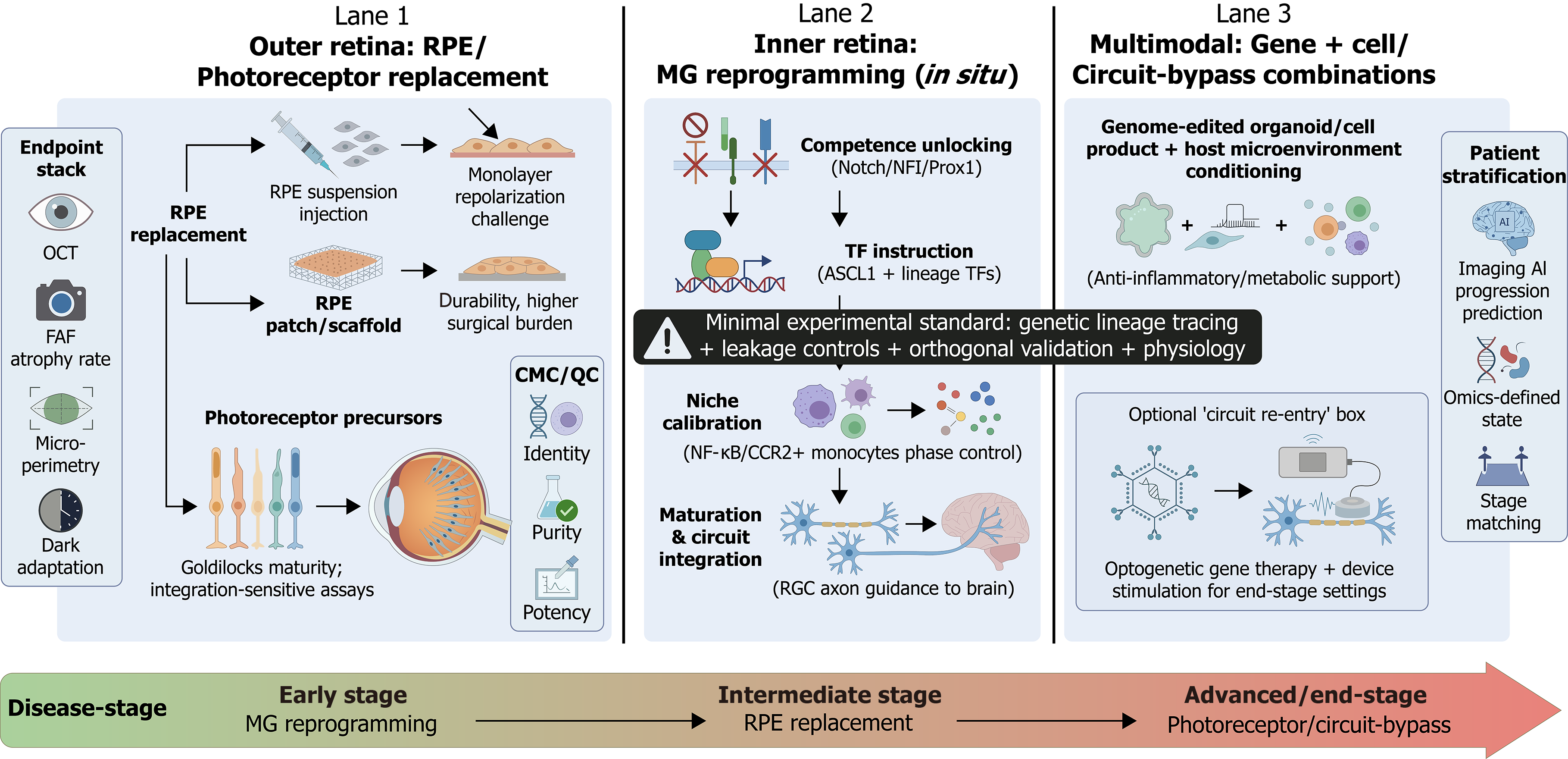
Figure 4 Regenerative ophthalmology strategies for retinal degeneration: Stage-matched, multimodal fate engineering.
This schematic synthesizes the clinical pathways for retinal fate engineering across three distinct therapeutic lanes, aligned with disease progression. Outer retina replacement: Strategies for RPE and photoreceptor replacement (discussed in “Regenerative strategy II: Clinical pathway and evidence hierarchy for cell-replacement therapy”), detailing delivery modalities (suspension vs scaffold) and the requirement for a multimodal endpoint stack (optical coherence tomography, fundus autofluorescence, microperimetry) alongside rigorous chemistry, manufacturing, and controls release criteria (identity, purity, potency) to ensure graft function (lane 1). Inner retina reprogramming: In situ Müller glia reprogramming (discussed in the section of Regenerative strategy III: MG reprogramming - from “feasible” to controllable, reproducible, and translatable) is depicted as a staged control sequence: Unlocking competence (e.g., via Notch/nuclear factor I inhibition), identifying lineage instruction (ASCL1), and calibrating the immune niche. A critical warning highlights the “minimal experimental standard” required to rule out viral leakage artifacts (lane 2). Multimodal combinations: “Gene + cell” and circuit-bypass strategies (discussed in “Gene + cell and multimodal combination strategies: Turning fate-control networks into therapies”) that couple biological replacement with host microenvironment conditioning or optogenetic re-entry (lane 3). Patient stratification: The timeline illustrates the necessity of precision stage-matching - deploying reprogramming for early-stage rescue, cell replacement for intermediate degeneration, and circuit-bypass for end-stage atrophy - guided by artificial intelligence-driven progression prediction (bottom). RPE: Retinal pigment epithelium; OCT: Optical coherence tomography; FAF: Fundus autofluorescence; CMC: Chemistry, manufacturing, and controls; QC: Quality control; MG: Müller glia; TF: Transcription factor; NFI: Nuclear factor I; Prox1: Prospero homeobox 1; NF-κB: Nuclear factor kappa B; CCR2+: C-C chemokine receptor type 2-positive; RGC: Retinal ganglion cell; AI: Artificial intelligence.
- Citation: Xie QQ, Zeng MQ, Mao LN, Han SJ, Sun D, Zheng ZG. Multilayered control of retinal stem/progenitor cell fate in the single-cell and organoid era: Developmental blueprints and regenerative opportunities. World J Stem Cells 2026; 18(4): 118621
- URL: https://www.wjgnet.com/1948-0210/full/v18/i4/118621.htm
- DOI: https://dx.doi.org/10.4252/wjsc.v18.i4.118621