
















Copyright: ©Author(s) 2026.
World J Stem Cells. Apr 26, 2026; 18(4): 118404
Published online Apr 26, 2026. doi: 10.4252/wjsc.v18.i4.118404
Published online Apr 26, 2026. doi: 10.4252/wjsc.v18.i4.118404
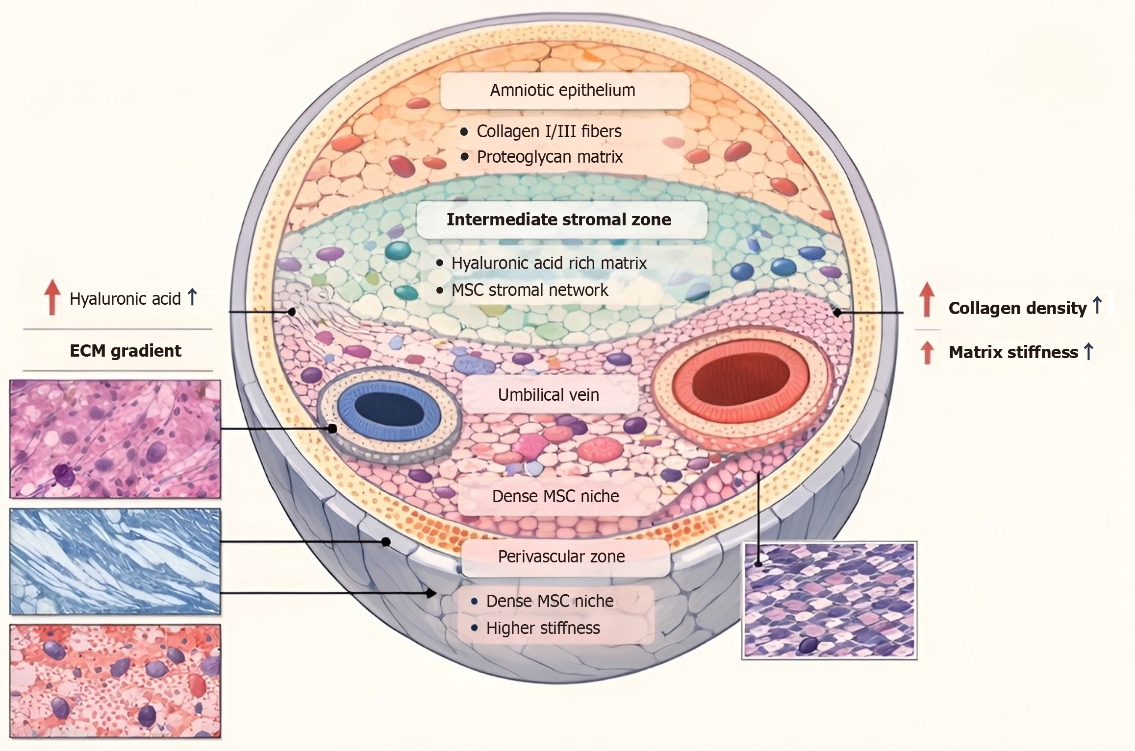
Figure 1 Cross-sectional view of the human umbilical cord.
Structural and cellular organization of Wharton’s jelly (WJ) within the human umbilical cord. The umbilical cord is composed of two arteries and one vein, and the whole is surrounded by WJ, with the latter covered by amniotic epithelium. The composition of WJ includes the subamniotic, intermediate stromal, and perivascular regions, each with distinct cell populations and biomechanical properties. The perivascular region is composed of more mesenchymal stem cells, resulting in the increased amount of collagen and the stiffness of the matrix, whereas the intermediate stromal region is composed mainly of a hyaluronic acid-based matrix. The histological images show the microstructural features of the matrix that contribute to the biomechanical properties of the WJ. ECM: Extracellular matrix; MSC: Mesenchymal stem cell.
- Citation: Habiba UE, Greene DL, Shamim S. Anatomy, function, biomechanics, and regenerative properties of Wharton’s jelly in the umbilical cord. World J Stem Cells 2026; 18(4): 118404
- URL: https://www.wjgnet.com/1948-0210/full/v18/i4/118404.htm
- DOI: https://dx.doi.org/10.4252/wjsc.v18.i4.118404