
















Copyright: ©Author(s) 2026.
World J Stem Cells. Apr 26, 2026; 18(4): 117652
Published online Apr 26, 2026. doi: 10.4252/wjsc.v18.i4.117652
Published online Apr 26, 2026. doi: 10.4252/wjsc.v18.i4.117652
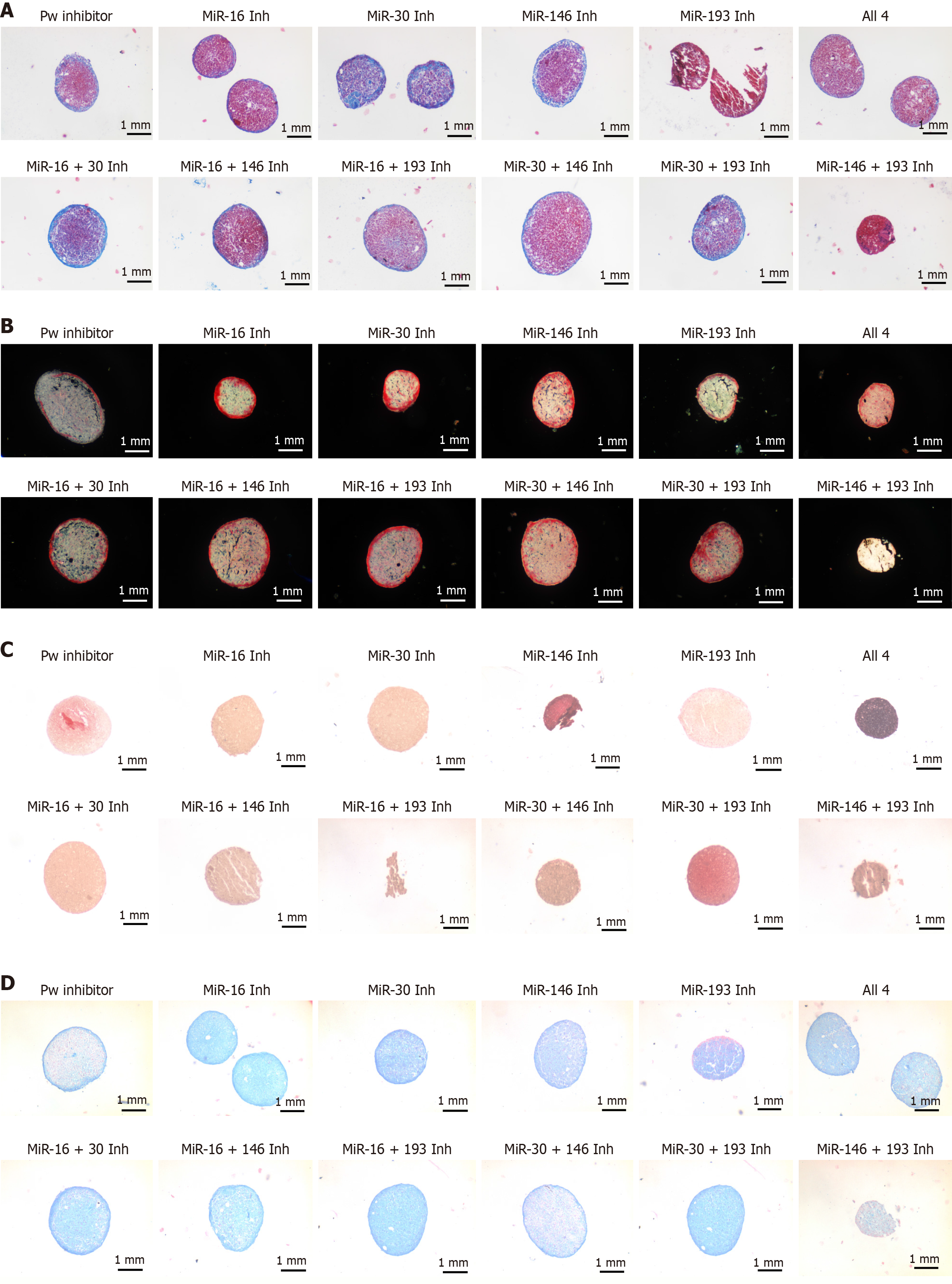
Figure 4 Histological assessment of cartilage-like matrix in micromasses at day 21.
A: Masson’s trichrome. Collagen appears blue, cytoplasm pink, and nuclei dark. Differences in collagen content and peripheral organisation are evident; B: Picrosirius Red under polarised light. Aggregated collagen fibrils show bright birefringence (red-orange), revealing variations in fibril abundance and orientation; C: Alcian Blue/Fast Red. Acidic sulphated mucopolysaccharides (sGAG) stain light blue; relative intensity reflects sGAG distribution across sections; D: Safranin O/Fast Green. sGAG/ proteoglycans stain dark orange-red, indicating cartilage-like matrix deposition. Representative images are shown for all experimental conditions; micrographs acquired at 40 × magnification.
- Citation: Fontiveros-Palomino M, Álvarez-Iglesias I, González-González A, Alfonso-Fernández A, Pérez-Campo FM. Modulation of osteoarthritis-related microRNAs using locked nucleic acid-antisense oligonucleotides boosts chondrogenic lineage commitment of mesenchymal progenitors. World J Stem Cells 2026; 18(4): 117652
- URL: https://www.wjgnet.com/1948-0210/full/v18/i4/117652.htm
- DOI: https://dx.doi.org/10.4252/wjsc.v18.i4.117652