
















Copyright: ©Author(s) 2026.
World J Stem Cells. Apr 26, 2026; 18(4): 115446
Published online Apr 26, 2026. doi: 10.4252/wjsc.v18.i4.115446
Published online Apr 26, 2026. doi: 10.4252/wjsc.v18.i4.115446
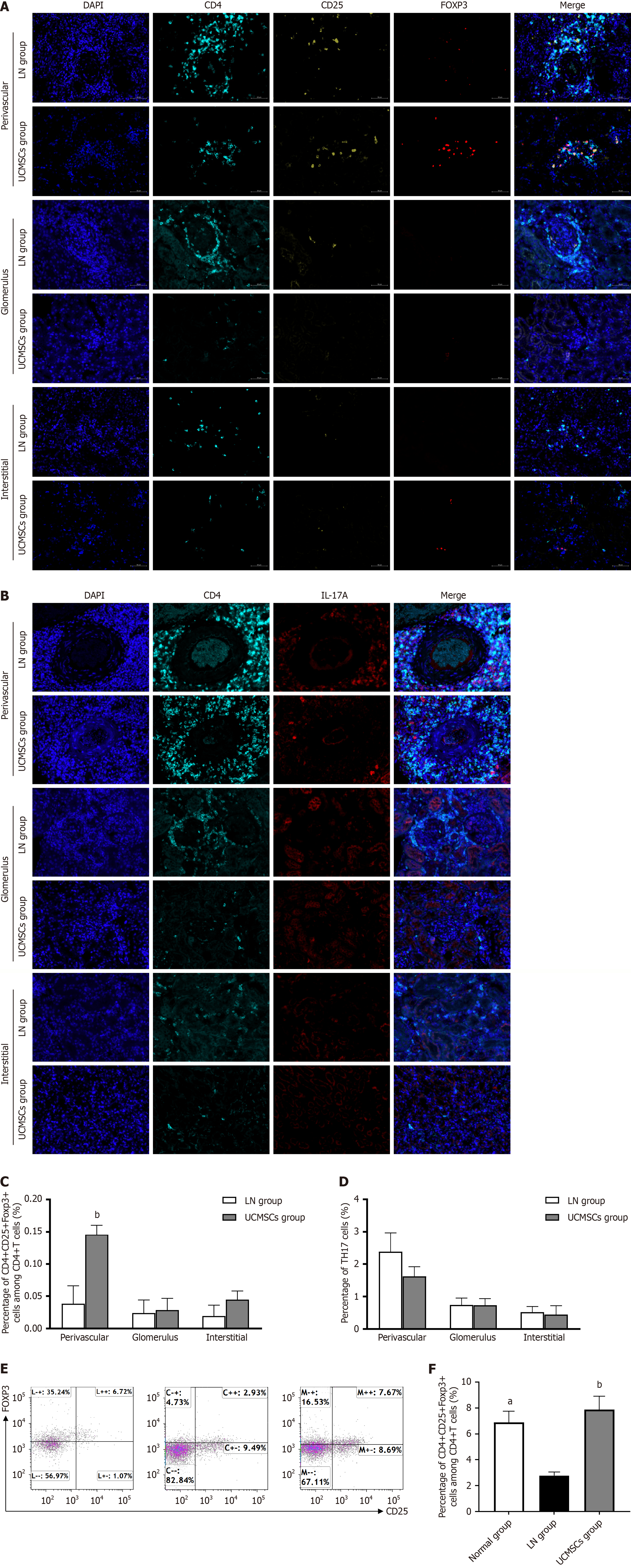
Figure 4 Changes in immune cells in the spleens and kidney tissues of MRL/Ipr mice.
A-D: Multiple immunofluorescence and quantitative analysis of regulatory T (Treg) (CD4+CD25+Foxp3+) (A and C); multiple immunofluorescence and quantitative analysis of T helper 17 (IL-17A+CD4+) (B and D); E: Flow cytometric analysis of the percentages of Treg (CD4+CD25+Foxp3+) in the spleen; F: Quantitative analysis of the percentages of Treg in the spleen. All data were expressed as mean ± SEM, aP < 0.05 vs lupus nephritis group, bP < 0.01 vs lupus nephritis group. LN: Lupus nephritis; UCMSCs: Umbilical cord mesenchymal stem cells; IL: Interleukin.
- Citation: He LJ, Meng MY, Huang CK, Liu SY, Wang P, Long W, Gao H, Yang LS, He S, He Y, Guo YF, Zhao YY, Liu Y, Hu LR, Li L, Hou ZL, Wang WJ, Wang XD. Mesenchymal stem cells alleviate renal injury of lupus nephritis mice by reconstructing the immune microenvironment of renal tissue. World J Stem Cells 2026; 18(4): 115446
- URL: https://www.wjgnet.com/1948-0210/full/v18/i4/115446.htm
- DOI: https://dx.doi.org/10.4252/wjsc.v18.i4.115446