
















Copyright: ©Author(s) 2026.
World J Stem Cells. Apr 26, 2026; 18(4): 115446
Published online Apr 26, 2026. doi: 10.4252/wjsc.v18.i4.115446
Published online Apr 26, 2026. doi: 10.4252/wjsc.v18.i4.115446
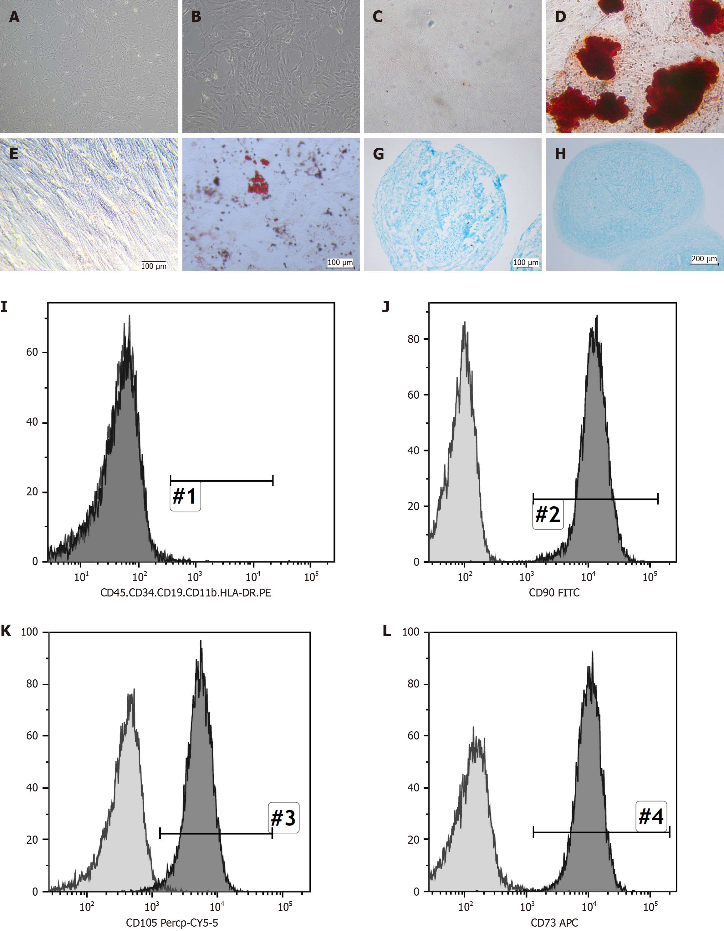
Figure 1 Umbilical cord mesenchymal stem cells morphology and identification.
A: Umbilical cord mesenchymal stem cells (UCMSCs) morphology is displayed at 50 × magnification; B: UCMSCs morphology is displayed at 200 × magnification; C and D: The effective differentiation of UCMSCs into osteoblasts was confirmed by alizarin red staining. At a magnification of 100 × representative pictures of the control group (C) and induction group (D) were captured; E and F: Oil Red O staining was used to successfully differentiate UCMSCs into adipocytes. At a magnification of 100 ×, representative pictures of the control group (E) and induction group (F) were captured; G and H: Alcian blue staining was used to identify the chondrocytes that were successfully differentiated from UCMSCs. At 200 × magnification, representative pictures of the control group (G) and induction group (H) were captured; I-L: Using flow cytometry, the surface markers of UCMSCs, such as CD90, CD105, CD73, CD45, CD34, CD11b, CD19, and HLA-DR, were examined. Of them, more than 99% of the cells were positive for CD90, CD105, and CD73. The other genes’ expression was nearly negligible.
- Citation: He LJ, Meng MY, Huang CK, Liu SY, Wang P, Long W, Gao H, Yang LS, He S, He Y, Guo YF, Zhao YY, Liu Y, Hu LR, Li L, Hou ZL, Wang WJ, Wang XD. Mesenchymal stem cells alleviate renal injury of lupus nephritis mice by reconstructing the immune microenvironment of renal tissue. World J Stem Cells 2026; 18(4): 115446
- URL: https://www.wjgnet.com/1948-0210/full/v18/i4/115446.htm
- DOI: https://dx.doi.org/10.4252/wjsc.v18.i4.115446