
















Copyright: ©Author(s) 2026.
World J Stem Cells. Apr 26, 2026; 18(4): 115218
Published online Apr 26, 2026. doi: 10.4252/wjsc.v18.i4.115218
Published online Apr 26, 2026. doi: 10.4252/wjsc.v18.i4.115218
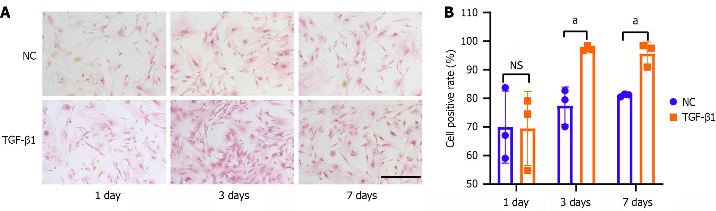
Figure 1 Transforming growth factor-β1 treatment induced collagen accumulation in tendon stem/progenitor cells.
A: Picro-Sirius red staining of tendon stem/progenitor cells after treatment with 5 ng/mL of transforming growth factor-β1 at 1 day, 3 days, and 7 days. Collagen is marked as red. Scale bars = 200 μm; B: The chart shows the percentage of the Picro-Sirius red-stained tendon stem/progenitor cells in different groups. All data are presented as the mean ± SD. aP < 0.05. NS: No significance; NC: Negative control; TGF-β1: Transforming growth factor-β1.
- Citation: Chen Y, Hu X, Jia XT, Gao RZ, Hou QS, Zhou SG. WTAP-mediated N6-methyladenosine modification of circ_PVT1 promotes proliferation and tenogenic differentiation of tendon stem/progenitor cells. World J Stem Cells 2026; 18(4): 115218
- URL: https://www.wjgnet.com/1948-0210/full/v18/i4/115218.htm
- DOI: https://dx.doi.org/10.4252/wjsc.v18.i4.115218