
















Copyright: ©Author(s) 2026.
World J Stem Cells. Mar 26, 2026; 18(3): 116388
Published online Mar 26, 2026. doi: 10.4252/wjsc.v18.i3.116388
Published online Mar 26, 2026. doi: 10.4252/wjsc.v18.i3.116388
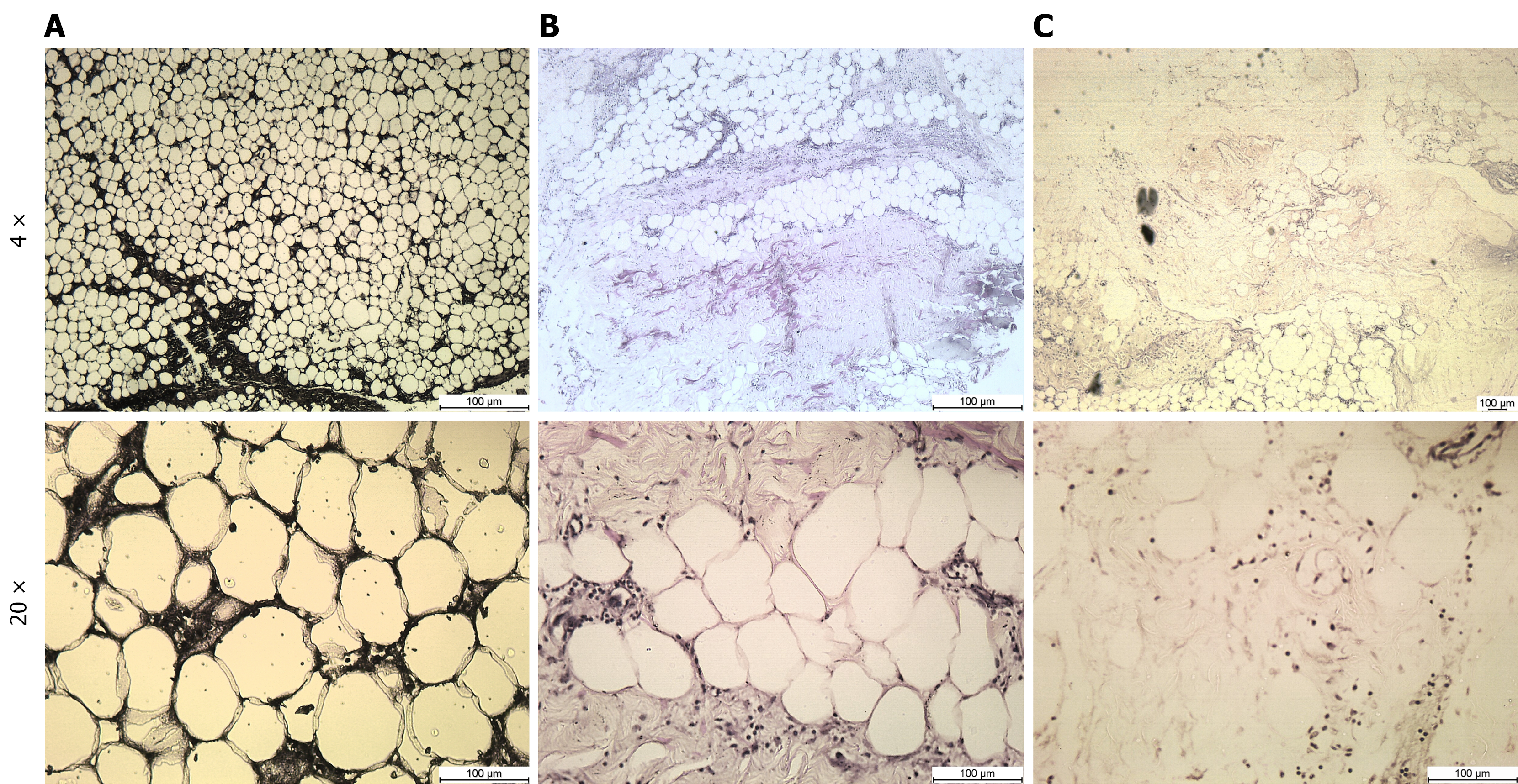
Figure 6 Histological evaluation of adipose tissue structure at different stages of collagenase digestion.
Representative photomicrographs of adipose tissue sections captured at 4 × (left) and 20 × (right) magnifications. A: Undigested adipose tissue showing a well-preserved and compact structure of densely packed, adipocytes with intact morphology and minimal extracellular matrix (ECM) disruption; B: Adipose tissue after the first round of collagenase digestion reveals partial ECM degradation and moderate adipocyte disruption, with some cells retaining morphology and others beginning to dissociate; C: Tissue after the second round of digestion demonstrates substantial structural breakdown, with complete loss of adipocyte integrity and prominent fibrous ECM remnants. This stage reflects advanced digestion and effective decellularization.
- Citation: Mushtaq A, Choudhery MS, Arif T, Niaz A, Pervaiz A. Maximizing pediatric stem cell harvest: A novel two-step digestion approach for adipose tissue. World J Stem Cells 2026; 18(3): 116388
- URL: https://www.wjgnet.com/1948-0210/full/v18/i3/116388.htm
- DOI: https://dx.doi.org/10.4252/wjsc.v18.i3.116388