
















Copyright: ©Author(s) 2026.
World J Stem Cells. Mar 26, 2026; 18(3): 116388
Published online Mar 26, 2026. doi: 10.4252/wjsc.v18.i3.116388
Published online Mar 26, 2026. doi: 10.4252/wjsc.v18.i3.116388
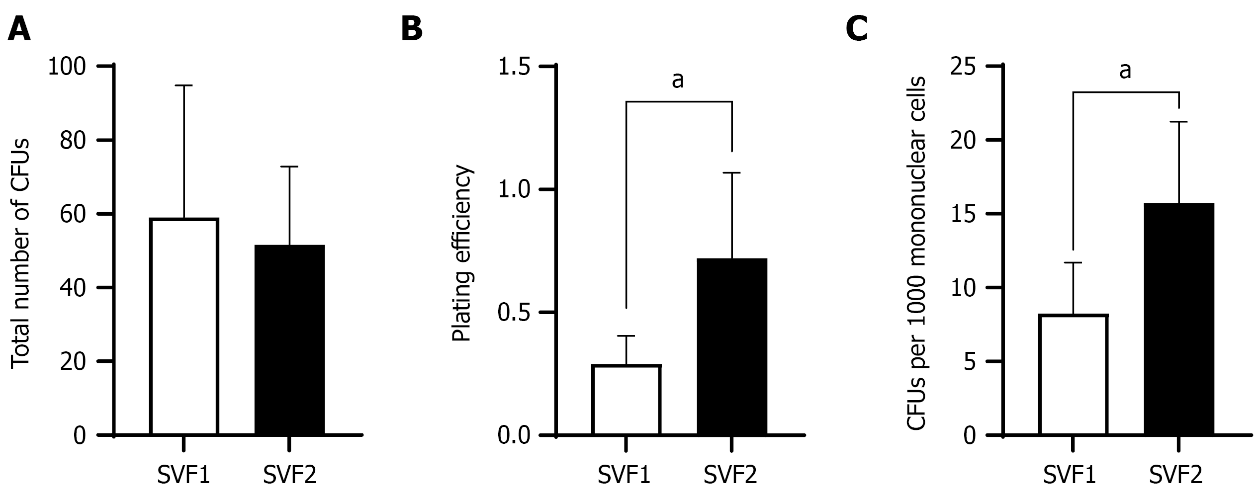
Figure 3 Comparison of colony forming units and clonogenic potential of stromal vascular fraction 1 and stromal vascular fraction 2.
A-C: Figure shows total number of colony forming units (CFU) (A), plating efficiency (B), and number of CFUs per 1000 mononucleated cells (C) in stromal vascular fraction 1 and stromal vascular fraction 2. Graphs were presented as mean ± SD and statistical significance was measured between stromal vascular fraction 1 and stromal vascular fraction 2 by unpaired two-tailed Student’s t-test. aP < 0.05. SVF: Stromal vascular fraction; CFU: Colony forming unit.
- Citation: Mushtaq A, Choudhery MS, Arif T, Niaz A, Pervaiz A. Maximizing pediatric stem cell harvest: A novel two-step digestion approach for adipose tissue. World J Stem Cells 2026; 18(3): 116388
- URL: https://www.wjgnet.com/1948-0210/full/v18/i3/116388.htm
- DOI: https://dx.doi.org/10.4252/wjsc.v18.i3.116388