
















Copyright: ©Author(s) 2026.
World J Stem Cells. Mar 26, 2026; 18(3): 115363
Published online Mar 26, 2026. doi: 10.4252/wjsc.v18.i3.115363
Published online Mar 26, 2026. doi: 10.4252/wjsc.v18.i3.115363
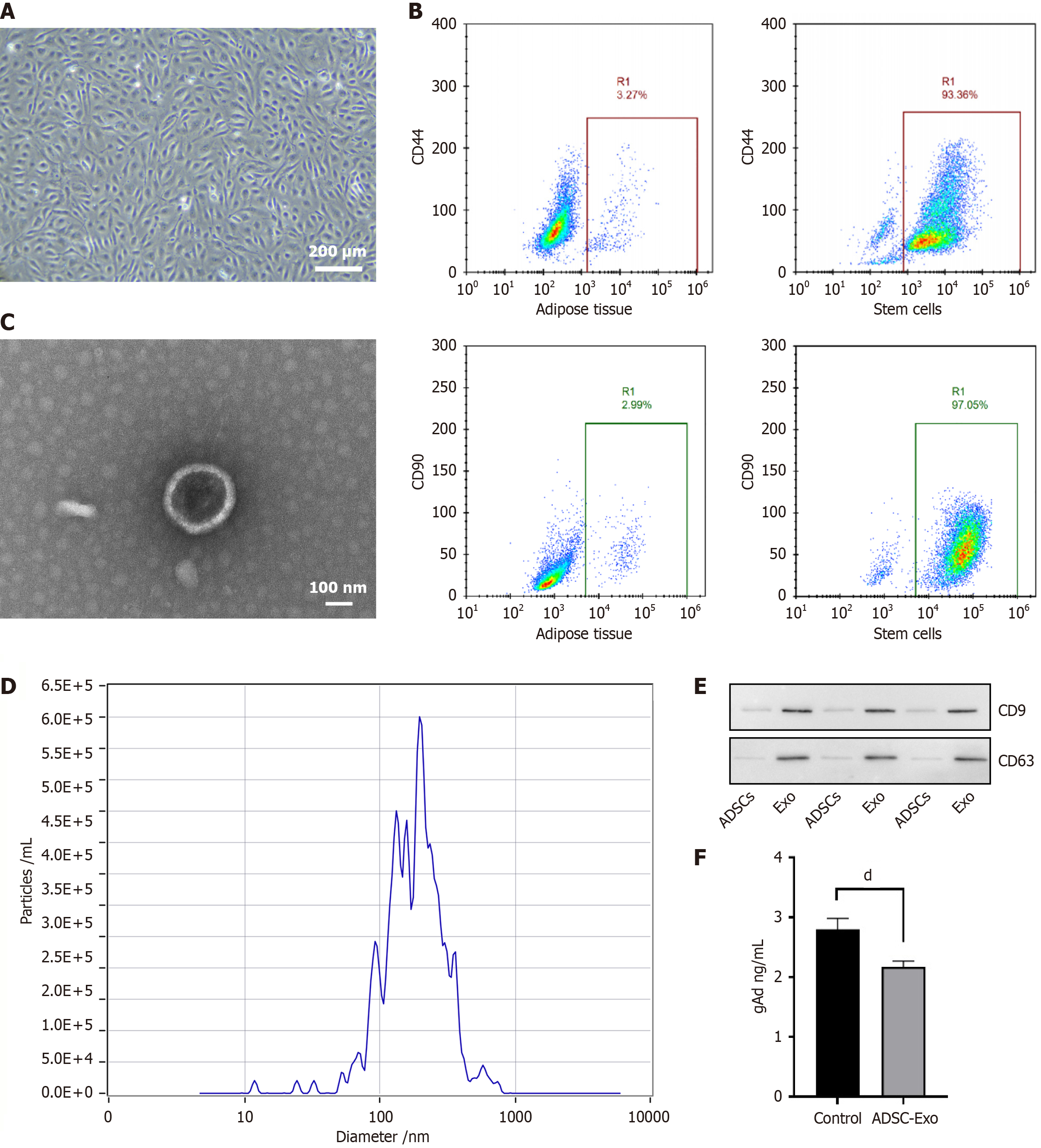
Figure 1 Isolation and characterization of rat mesenchymal stem cells and adipose-derived mesenchymal stem cells-derived exosomes.
A: Photomicrographic image of adipose-derived mesenchymal stem cells (ADSCs), scale bars = 200 μm; B: Flow cytometry analysis of the ADSCs revealed that they expressed a high cluster of CD44 and CD90; C: The ADSCs-derived exosomes appeared double-layered membranous under transmission electron microscopy, scale bars = 100 nm; D: Size distribution of ADSCs-derived exosomes by nanoparticle tracking analysis; E: Western blots exosome markers CD9 and CD63; F: Assessment of exosome loading efficiency (dP < 0.0001). ADSCs: Adipose-derived mesenchymal stem cells; Exo: Exosome; gAd: Globular adiponectin.
- Citation: Wang AR, Zhang YY, Liu YJ, Ren LN, Yan QH, Wang KF, Wang Y. Exosomes of adipose-derived mesenchymal stem cells loaded with globular adiponectin improve islet function for type 2 diabetes. World J Stem Cells 2026; 18(3): 115363
- URL: https://www.wjgnet.com/1948-0210/full/v18/i3/115363.htm
- DOI: https://dx.doi.org/10.4252/wjsc.v18.i3.115363