
















Copyright: ©Author(s) 2026.
World J Stem Cells. Mar 26, 2026; 18(3): 114746
Published online Mar 26, 2026. doi: 10.4252/wjsc.v18.i3.114746
Published online Mar 26, 2026. doi: 10.4252/wjsc.v18.i3.114746
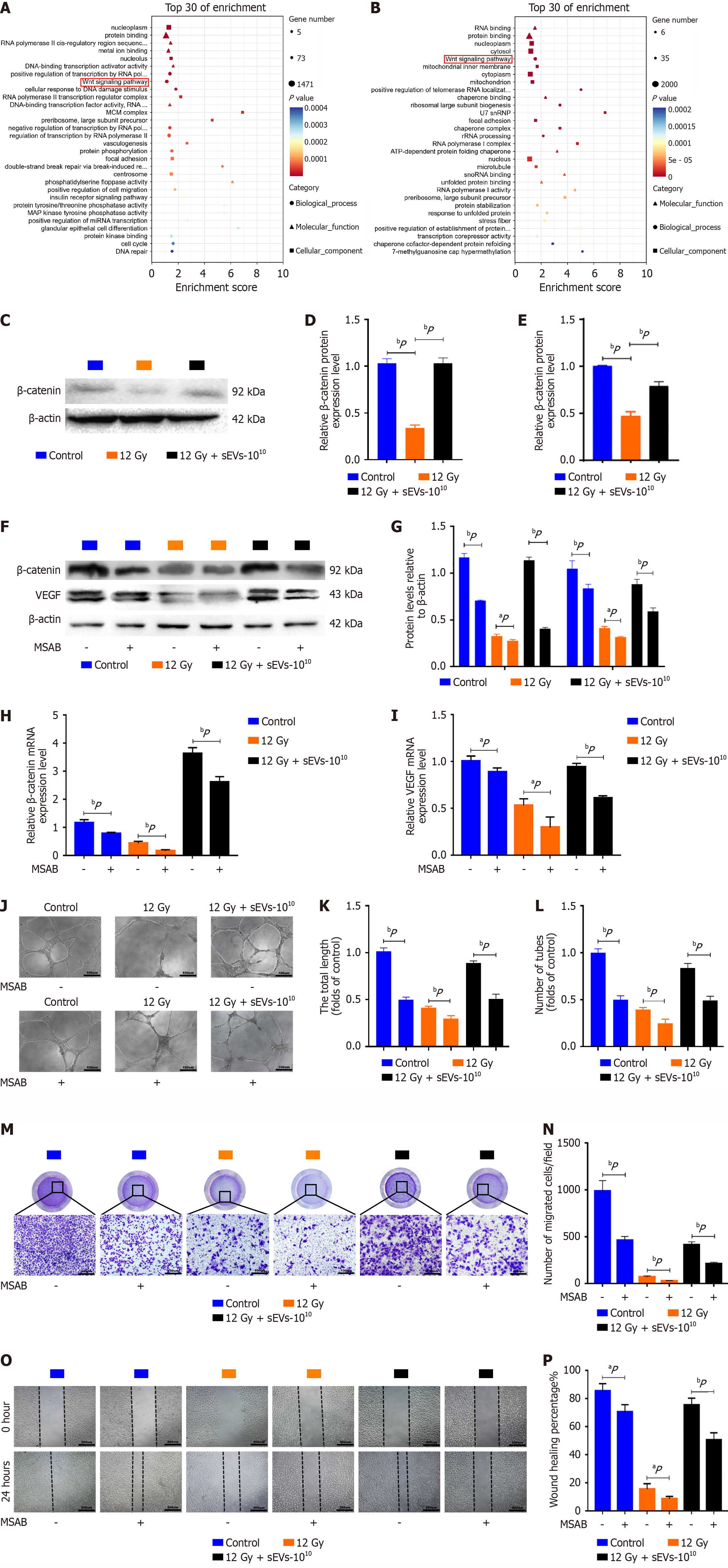
Figure 6 Human amniotic mesenchymal stem cell-derived small extracellular vesicles activated Wnt/β-catenin expression in irradiated human umbilical vein endothelial cells.
A: Kyoto Encyclopedia of Genes and Genomes pathway analysis illustrating the top 30 downregulated genes in human umbilical vein endothelial cells (HUVECs) between the 12 Gy and control groups; B: Kyoto Encyclopedia of Genes and Genomes pathway analysis illustrating the top 30 upregulated genes in HUVECs between the 12 Gy + 1010 human amniotic mesenchymal stem cell-derived small extracellular vesicles (hAMSCs-sEVs) and 12 Gy groups; C-E: Western blot and polymerase chain reaction analysis of β-catenin expression in HUVECs in the control, 12 Gy and 12 Gy + 1010 hAMSCs-sEVs groups; F-I: Western blot and polymerase chain reaction analysis of β-catenin and vascular endothelial growth factor expression in HUVECs in the control, 12 Gy and 12 Gy + 1010 hAMSCs-sEVs groups treated with or without the selective inhibitor MSAB; J-L: Tube formation assay in the control, 12 Gy and 12 Gy + 1010 hAMSCs-sEVs groups treated with or without the selective inhibitor MSAB. Scale bar = 100 μm; M and N: Transwell assay in the control, 12 Gy and 12 Gy + 1010 hAMSCs-sEVs groups treated with or without the selective inhibitor MSAB. Scale bar = 100 μm; O and P: Scratch wound healing assay in the control, 12 Gy and 12 Gy + 1010 hAMSCs-sEVs groups treated with or without the selective inhibitor MSAB. aP < 0.05, bP < 0.01. VEGF: Vascular endothelial growth factor; sEVs: Small extracellular vesicles.
- Citation: Zhou HF, Xu JM, Li J, Xu F, Li JF, Yu Y, Yuan H, Bian YF, Wang YL, Du YF. Human amniotic mesenchymal stem cell-derived small extracellular vesicles repair of jaw osteoradionecrosis. World J Stem Cells 2026; 18(3): 114746
- URL: https://www.wjgnet.com/1948-0210/full/v18/i3/114746.htm
- DOI: https://dx.doi.org/10.4252/wjsc.v18.i3.114746