
















Copyright: ©Author(s) 2026.
World J Stem Cells. Mar 26, 2026; 18(3): 113900
Published online Mar 26, 2026. doi: 10.4252/wjsc.v18.i3.113900
Published online Mar 26, 2026. doi: 10.4252/wjsc.v18.i3.113900
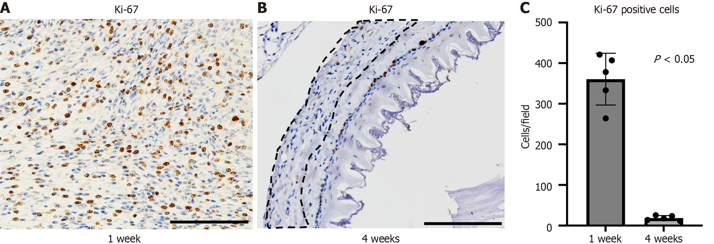
Figure 6 Ki-67-positive cells in the transplanted myoblast cell sheets at each time point.
A and B: Ki-67 immunostaining (× 400, scale bar: 100 μm) of the transplantation group at 1 week postoperatively showed that the layer formed by the myoblast cell sheet contained numerous Ki-67-positive cells (A), and 4 weeks postoperatively showed very few Ki-67 positive cells (proliferating cells) in the transplanted myoblast sheet layer (enclosed by a dotted line) (B); C: The number of Ki-67-positive cells per high-power field (× 400) detected via Ki-67 immunostaining showed that Ki-67-positive cells in the myoblast sheet-transplanted area at 4 weeks post-transplantation were significantly reduced compared to that at 1 week post-transplantation.
- Citation: Enjoji T, Kobayashi S, Miyamoto D, Ogawa S, Tetsuo H, Soyama A, Adachi T, Eguchi S, Kanetaka K. Allogeneic myoblast cell sheet transplantation for esophageal wall reinforcement. World J Stem Cells 2026; 18(3): 113900
- URL: https://www.wjgnet.com/1948-0210/full/v18/i3/113900.htm
- DOI: https://dx.doi.org/10.4252/wjsc.v18.i3.113900