
















©Author(s) (or their employer(s)) 2026.
World J Stem Cells. Feb 26, 2026; 18(2): 114032
Published online Feb 26, 2026. doi: 10.4252/wjsc.v18.i2.114032
Published online Feb 26, 2026. doi: 10.4252/wjsc.v18.i2.114032
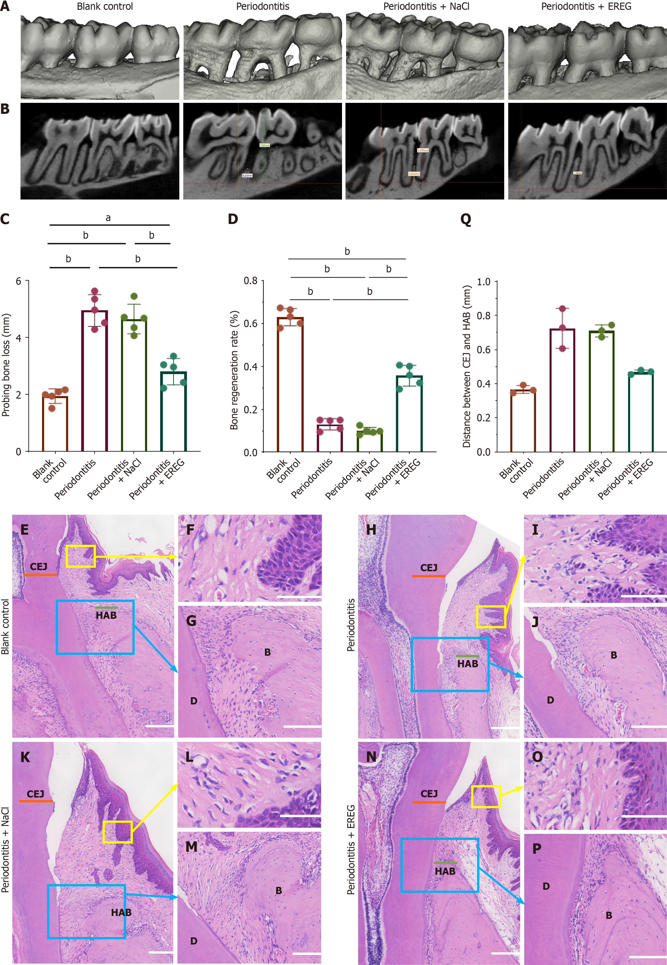
Figure 4 Local injection of epiregulin promoted the regeneration of periodontal tissue in a rat model of periodontitis.
A: Three-dimensional micro-computed tomography reconstruction of mouse maxillary from the three groups; B: Sagittal-sectional images of maxillary; C and D: Micro-computed tomography measurement of bone formation (n = 5, one-way analysis of variance); E-M: Hematoxylin and eosin staining indicated new periodontal tissue regeneration from coronal-sectional images of maxillary in the blank control group (E), periodontitis group (H), periodontitis + sodium chloride (NaCl) (K) and periodontitis + epiregulin (EREG) (N). Scale bar: 200 μm; hematoxylin and eosin staining indicated Sharpey’s fibers in the blank control group (G), periodontitis group (J), periodontitis + NaCl (M) and periodontitis + EREG (P). Scale bar: 100 μm; hematoxylin and eosin staining indicated inflammatory cells infiltration in the blank control group (F), periodontitis group (I), periodontitis + NaCl (L) and periodontitis + EREG (O). Scale bar: 50 μm; Q: Quantitative analysis of distance between cemento-enamel junction (CEJ) and height of alveolar bone (n = 3, Kruskal-Wallis test). Error bars represent the standard deviation. aP ≤ 0.05; bP ≤ 0.01. B: Bone; D: Dentin; HAB: Height of alveolar bone.
- Citation: Zhao YC, Li GY, Chen S, Wang YL, Feng JY, Cao Y. Epiregulin enhances periodontal tissue regeneration by promoting bone marrow mesenchymal stem cell functions under inflammatory niches. World J Stem Cells 2026; 18(2): 114032
- URL: https://www.wjgnet.com/1948-0210/full/v18/i2/114032.htm
- DOI: https://dx.doi.org/10.4252/wjsc.v18.i2.114032