
















©Author(s) (or their employer(s)) 2026.
World J Stem Cells. Feb 26, 2026; 18(2): 113694
Published online Feb 26, 2026. doi: 10.4252/wjsc.v18.i2.113694
Published online Feb 26, 2026. doi: 10.4252/wjsc.v18.i2.113694
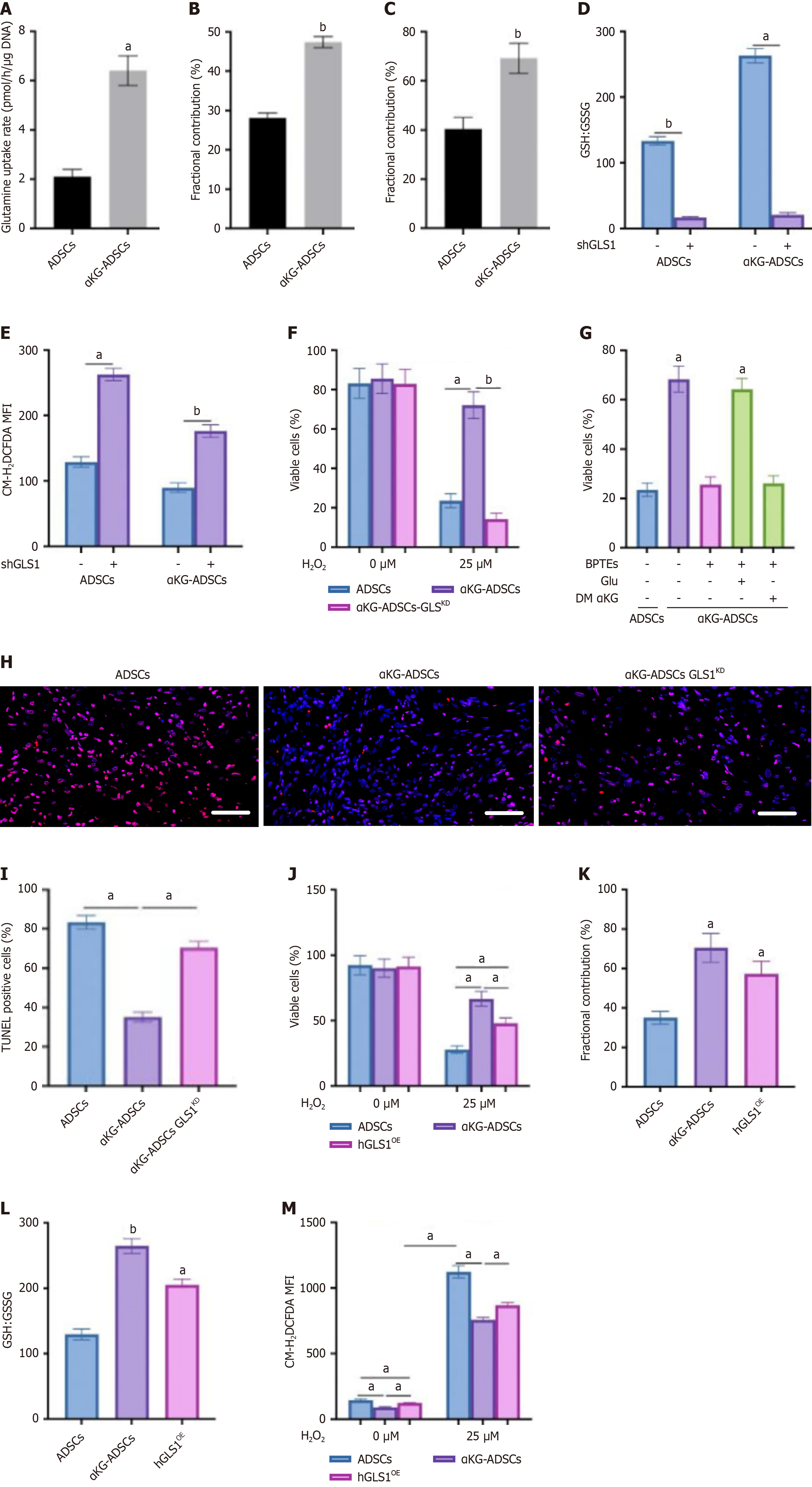
Figure 3 GLS1 regulates glutamine-mediated reactive oxygen species scavenging in adipose-derived stem cells treated with α-ketoglutarate and contributes to cell survival.
A: Glutamine uptake rate in mouse adipose-derived stem cells (ADSCs) and α-ketoglutarate-ADSCs (αKG-ADSCs); B and C: Fractional contribution of [U-13C]-glutamine to glutamate (B) and glutathione (GSH) (C) in mouse ADSCs and αKG-ADSCs; D and E: GSH to GSSG disulfide levels (D) and total intracellular reactive oxygen species levels (E) in mouse ADSCs and αKG-ADSCs after genetic silencing of GLS1 [scrambled shRNA(-) or shGLS1 (+)]; F: Cell viability of mouse ADSCs and αKG-ADSCs after treatment with 25 μmol/L H2O2. αKG-ADSCs-GLSKD are αKG-ADSCs transduced with shGLS1; G: Rescue of cell viability of BPTES-treated αKG-ADSCs cells during oxidative stress (25 μmol/L H2O2) with glutamate, but not with dimethyl α-ketoglutarate; H and I: Terminal deoxynucleotidyl transferase dUTP nick end labeling immunostaining (H) of the mouse burn wounds treated with ADSCs, αKG-ADSCs, and ADSCs-GLSKD, with quantification (I); J: Cell viability of ADSCs, αKG-ADSCs, and hGLS1-overexpressing (hGLS1OE) αKG-ADSCs after treatment with 25 μmol/L H2O2; K: Fractional contribution of [U-13C]-glutamine to GSH; L: Ratio of GSH to GSH disulfide; M: Total intracellular reactive oxygen species levels with or without H2O2 treatment. Data are presented with means ± SEM (aP < 0.05, bP < 0.01). ADSCs: Adipose-derived stem cells; αKG-ADSCs: Alpha-ketoglutarate adipose-derived stem cells; GSH: Glutathione; GSSG: Glutathione disulfide; MFI: Mean fluorescent intensity; DM-αKG: Dimethyl α-ketoglutarate; TUNEL: Terminal deoxynucleotidyl transferase dUTP nick end labeling.
- Citation: Dilimulati D, Dilimulati D, Cui L. Alpha-ketoglutarate enhances adipose-derived stem cells survival in wound healing by hypoxia-inducible factor 1-alpha-mediated redox homeostasis and glycogen-dependent bioenergetics. World J Stem Cells 2026; 18(2): 113694
- URL: https://www.wjgnet.com/1948-0210/full/v18/i2/113694.htm
- DOI: https://dx.doi.org/10.4252/wjsc.v18.i2.113694