
















©Author(s) (or their employer(s)) 2026.
World J Stem Cells. Feb 26, 2026; 18(2): 112940
Published online Feb 26, 2026. doi: 10.4252/wjsc.v18.i2.112940
Published online Feb 26, 2026. doi: 10.4252/wjsc.v18.i2.112940
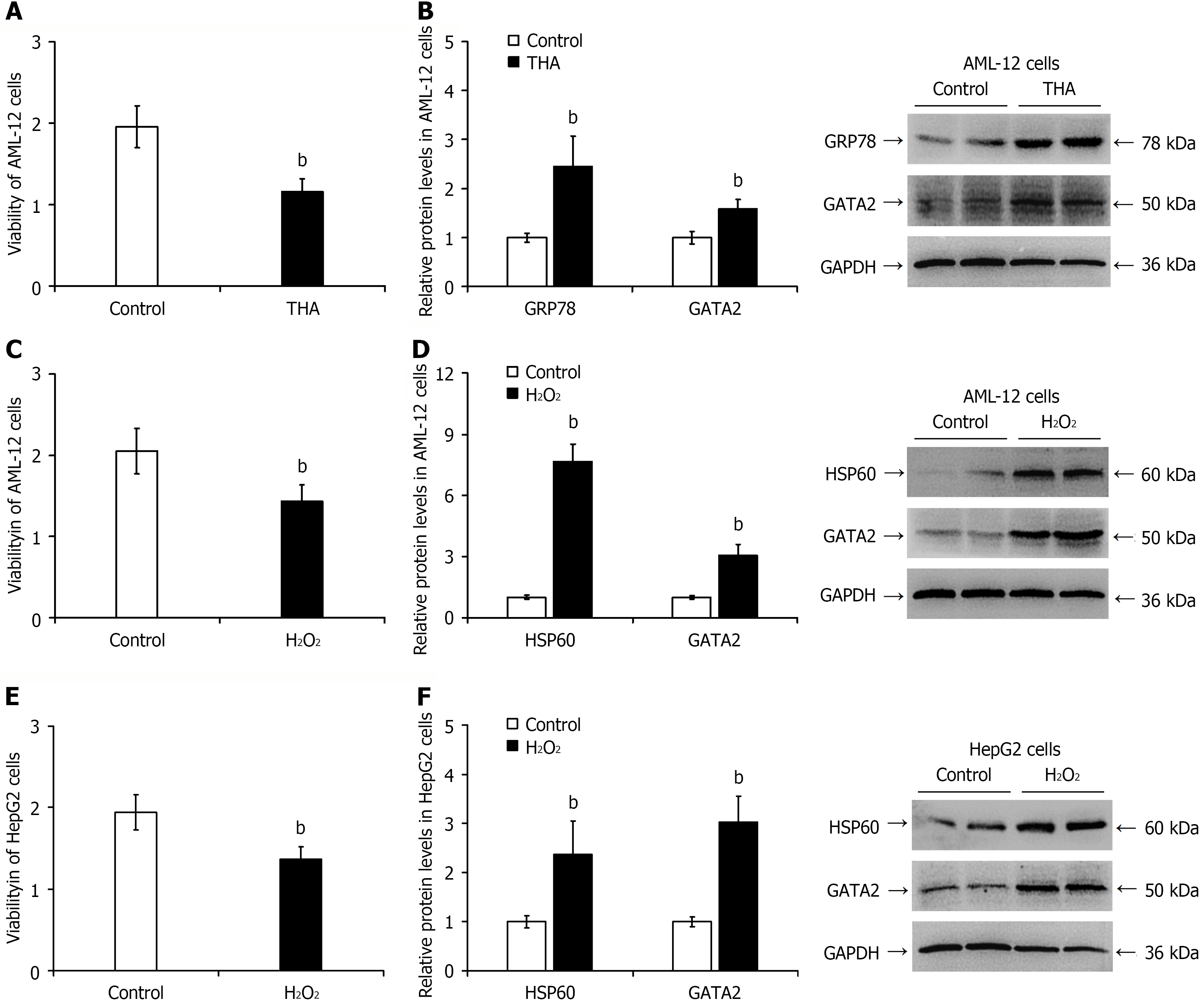
Figure 6 Elevated GATA2 protein expression in hepatocytes under endoplasmic reticulum stress and oxidative stress.
A: Cell viability assessed by Cell Counting Kit-8 assay in thapsigargin-treated AML-12 cells; B: Semi-quantitative analysis of thapsigargin-induced GATA2 protein expression by western blotting (representative blots shown); C and E: Cell viability measured by Cell Counting Kit-8 assay in AML-12 (C) and HepG2 cells (E) treated with H2O2; D and F: Semi-quantitative analysis of H2O2-induced GATA2 protein expression by western blotting in AML-12 (D) and HepG2 cells (F) (representative blots shown). bP < 0.01 vs control group. HSP60: Heat shock protein 60.
- Citation: Chen YF, Li SQ, Zhang J, Ma WT, Zhou Y, Rao JX, Yi Y, Cheng QJ, Zhong WW, Chen H, Chen YH, Luo YW, He YH. GATA2 deficiency exacerbates chronic liver injury via disrupting hepatocyte death-regeneration balance: Clinical, histopathological, and molecular evidence. World J Stem Cells 2026; 18(2): 112940
- URL: https://www.wjgnet.com/1948-0210/full/v18/i2/112940.htm
- DOI: https://dx.doi.org/10.4252/wjsc.v18.i2.112940