
















©Author(s) (or their employer(s)) 2026.
World J Stem Cells. Feb 26, 2026; 18(2): 112940
Published online Feb 26, 2026. doi: 10.4252/wjsc.v18.i2.112940
Published online Feb 26, 2026. doi: 10.4252/wjsc.v18.i2.112940
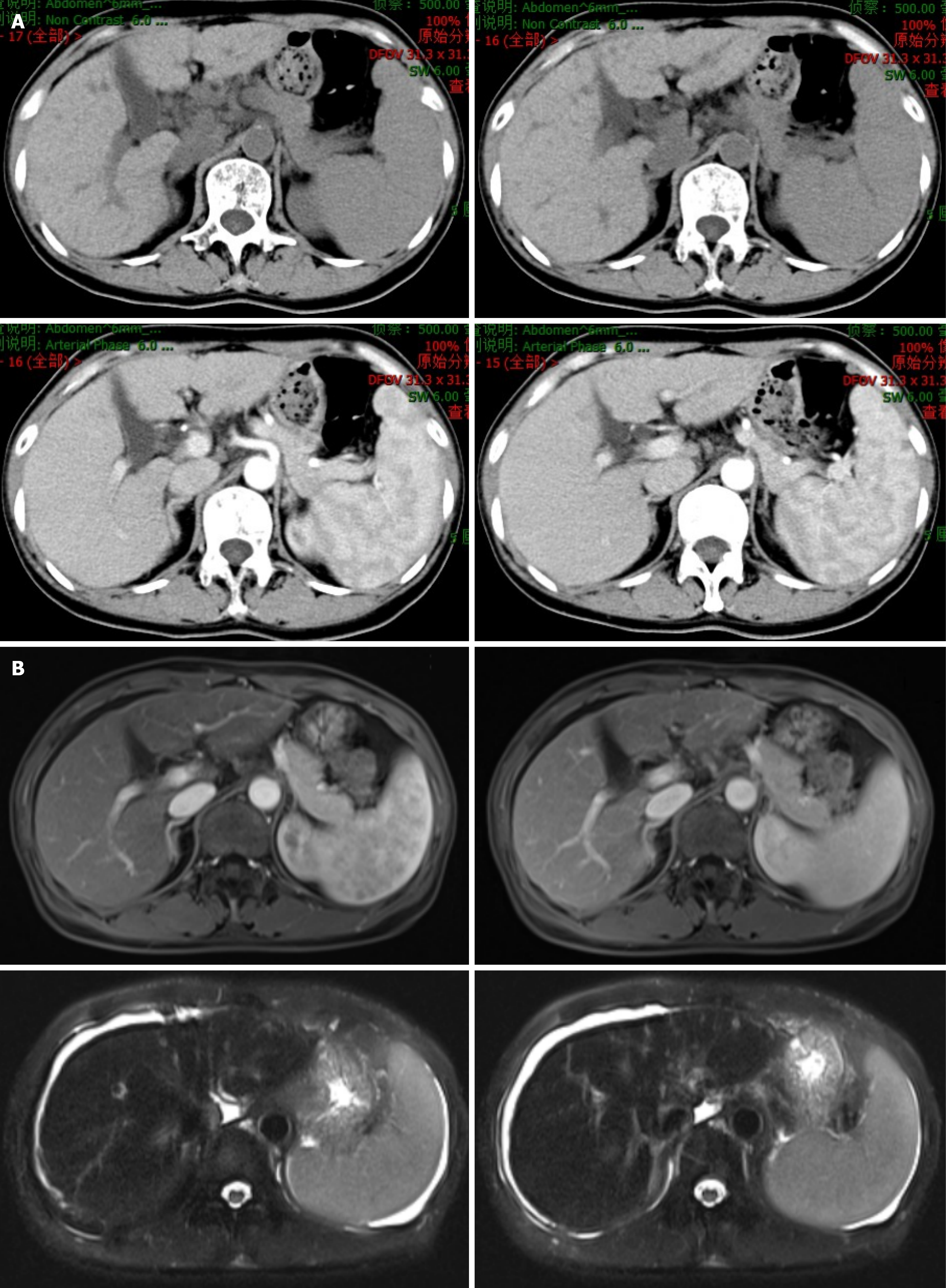
Figure 3 Abdominal magnetic resonance imaging and computed tomography findings of cirrhosis during follow-up.
A: Upper abdominal computed tomography (plain and enhanced) scans revealed irregular margins with slightly widened fissures, and strip low-density shadows around the portal vein in the liver; B: Upper abdominal magnetic resonance imaging revealed heterogeneous signal intensity with an irregular contour, slightly widened fissures, heterogeneous enhancement, and a long T2 signal around the portal vein in the liver. The spleen was enlarged. The upper abdominal magnetic resonance imaging findings indicated cirrhosis with splenomegaly and ascites. No abnormal enhancement was observed on the enhanced scan. The spleen was enlarged.
- Citation: Chen YF, Li SQ, Zhang J, Ma WT, Zhou Y, Rao JX, Yi Y, Cheng QJ, Zhong WW, Chen H, Chen YH, Luo YW, He YH. GATA2 deficiency exacerbates chronic liver injury via disrupting hepatocyte death-regeneration balance: Clinical, histopathological, and molecular evidence. World J Stem Cells 2026; 18(2): 112940
- URL: https://www.wjgnet.com/1948-0210/full/v18/i2/112940.htm
- DOI: https://dx.doi.org/10.4252/wjsc.v18.i2.112940