
















©Author(s) (or their employer(s)) 2026.
World J Stem Cells. Feb 26, 2026; 18(2): 112940
Published online Feb 26, 2026. doi: 10.4252/wjsc.v18.i2.112940
Published online Feb 26, 2026. doi: 10.4252/wjsc.v18.i2.112940
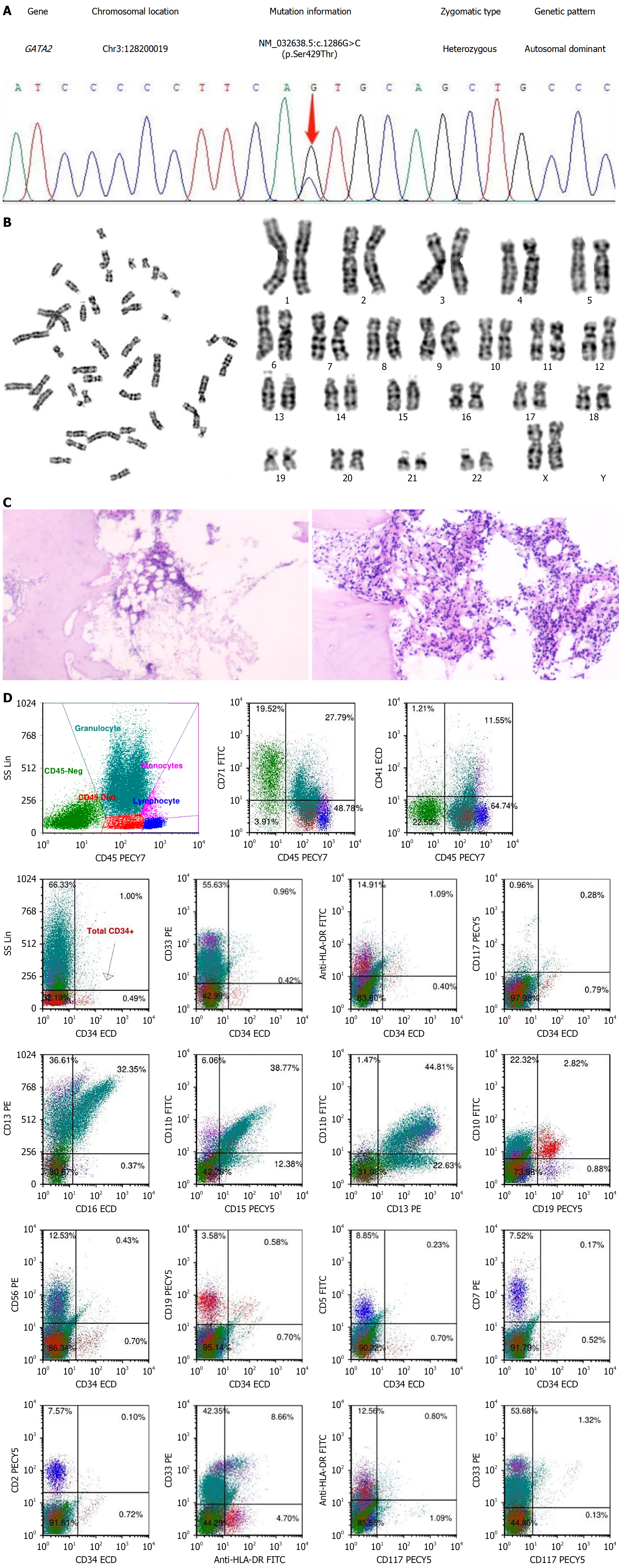
Figure 2 Identification of GATA2 mutation and diagnosis of aplastic anemia in the patient.
A: Sanger sequencing of the gene; B: Karyotype analysis; C: Pathological findings of bone marrow biopsy. The proliferation of bone marrow nucleated cells was low; the erythroid lineage was mainly composed of intermediate and late-stage cells; megakaryocytes were rare; lymphocytes were scattered in small numbers; CD34 small vessels (+); few CD61 megakaryocytes (+); D: Flow cytometric analysis. The proportion of CD34+ cells in nuclear cells was about 0.49%, with no obvious abnormality in the immunophenotype. The relative proportion of granulocytes was normal, and the immunophenotypes CD13, CD16, CD15, and CD11b were disordered. Approximately 2.60% of CD19+CD10+ immature B lymphocytes, consistent with normal B progenitor cell proliferation.
- Citation: Chen YF, Li SQ, Zhang J, Ma WT, Zhou Y, Rao JX, Yi Y, Cheng QJ, Zhong WW, Chen H, Chen YH, Luo YW, He YH. GATA2 deficiency exacerbates chronic liver injury via disrupting hepatocyte death-regeneration balance: Clinical, histopathological, and molecular evidence. World J Stem Cells 2026; 18(2): 112940
- URL: https://www.wjgnet.com/1948-0210/full/v18/i2/112940.htm
- DOI: https://dx.doi.org/10.4252/wjsc.v18.i2.112940