
















©Author(s) (or their employer(s)) 2026.
World J Stem Cells. Feb 26, 2026; 18(2): 112940
Published online Feb 26, 2026. doi: 10.4252/wjsc.v18.i2.112940
Published online Feb 26, 2026. doi: 10.4252/wjsc.v18.i2.112940
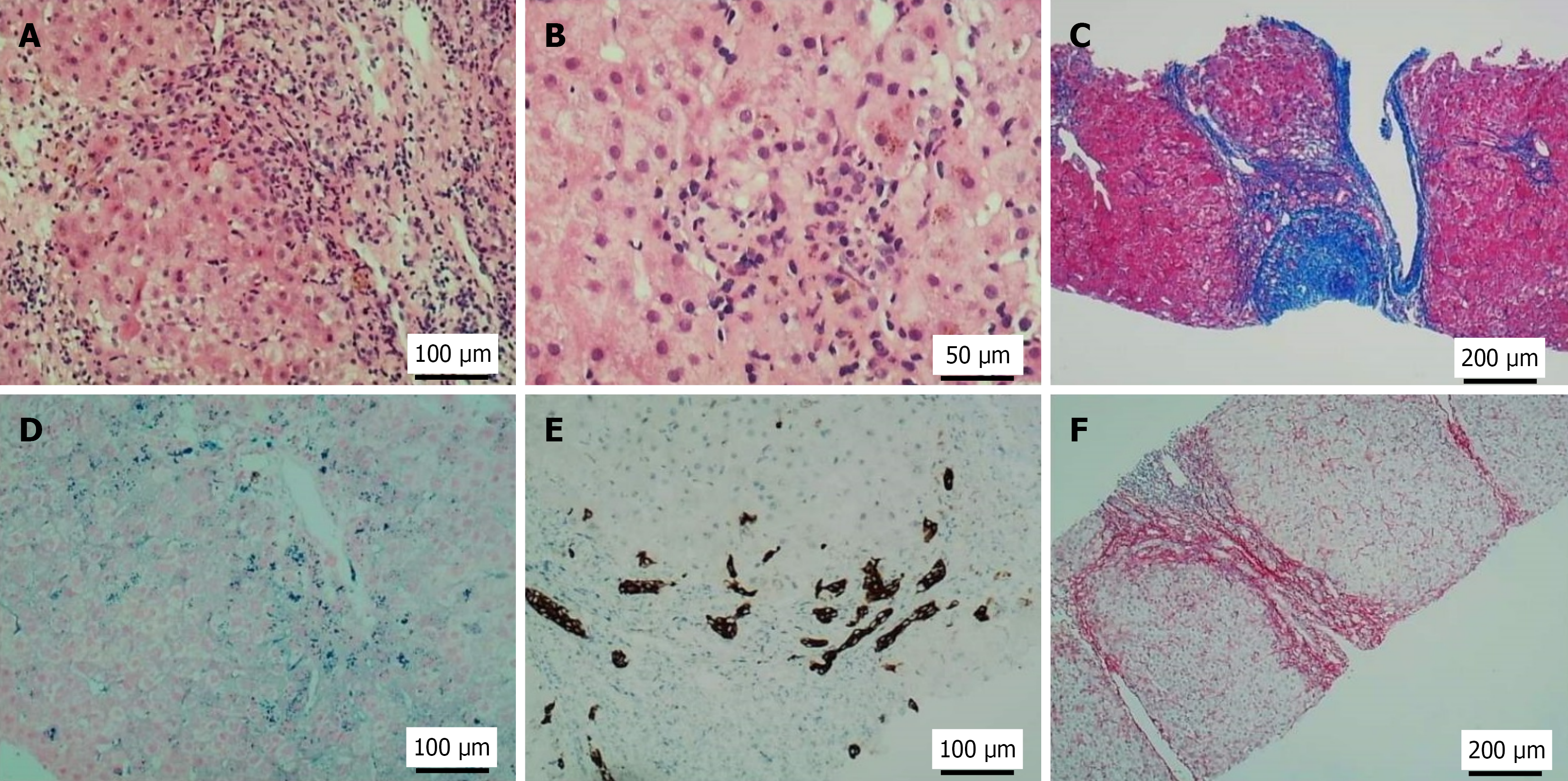
Figure 1 Pathological findings of liver biopsy in the patient.
A: Moderate interface inflammation with infiltration of lymphocytes and neutrophils; B: Hepatocytes containing yellow-brown granules and focal confluent necrosis; C: Masson trichrome stain: Portal-based fibrous expansion and septum (blue collagen); D: Prussian blue stain: Iron granules (blue-black deposits); E: Cytokeratin 7 immunohistochemistry: Biliary differentiation; F: Sirius red stain: Pericellular/periportal fibrosis (red/orange birefringence).
- Citation: Chen YF, Li SQ, Zhang J, Ma WT, Zhou Y, Rao JX, Yi Y, Cheng QJ, Zhong WW, Chen H, Chen YH, Luo YW, He YH. GATA2 deficiency exacerbates chronic liver injury via disrupting hepatocyte death-regeneration balance: Clinical, histopathological, and molecular evidence. World J Stem Cells 2026; 18(2): 112940
- URL: https://www.wjgnet.com/1948-0210/full/v18/i2/112940.htm
- DOI: https://dx.doi.org/10.4252/wjsc.v18.i2.112940