
















Copyright: ©Author(s) 2026.
World J Stem Cells. Oct 26, 2026; 18(10): 125852
Published online Oct 26, 2026. doi: 10.4252/wjsc.125852
Published online Oct 26, 2026. doi: 10.4252/wjsc.125852
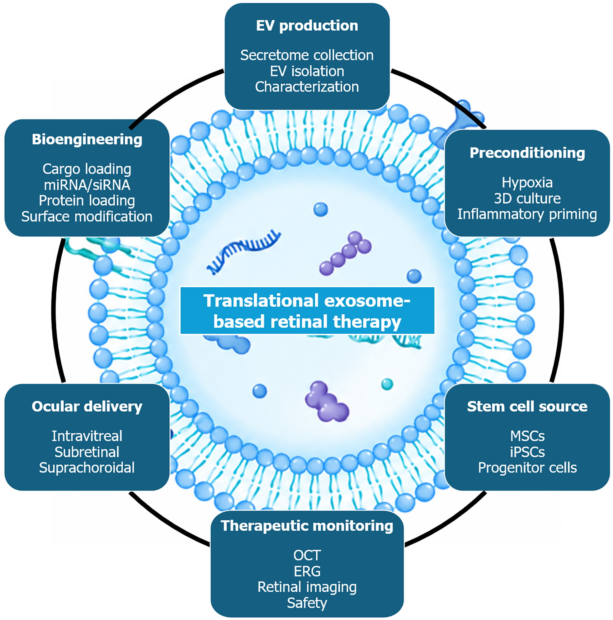
Figure 2 Proposed conceptual translational framework for bioengineered extracellular vesicle-based therapies in retinal disease.
The proposed workflow begins with the selection of an appropriate stem cell source. Viable sources include bone marrow-, adipose tissue-, umbilical cord-, Wharton’s jelly-derived mesenchymal stem cells; embryonic stem cells; induced pluripotent stem cells (iPSCs); neural or retinal progenitor cells; and patient-derived iPSC-derived retinal cells. Before secretome extraction and extracellular vesicle (EV) isolation, parental cells may be exposed to hypoxic or inflammatory stimuli, cultured in three-dimensional systems, or genetically modified to enhance the therapeutic properties of resultant vesicles. EV preparations should then undergo product-specific quality control including particle size and abundance, protein concentration, EV-associated markers, assessment of non-EV contaminants, cargo composition, biological potency, batch-to-batch consistency, and stability. Particle abundance and protein mass should be reported as distinct, non-interchangeable metrics. Bioengineering may be introduced either upstream through modification of the parental cell; or downstream through RNA, protein, or drug loading, surface functionalization, ligand-mediated targeting, and the development of hybrid vesicle systems. Engineered vesicles may be delivered via either intravitreal, subretinal, suprachoroidal administration; or incorporated into biomaterial-based delivery platforms. Potential targets include photoreceptors, retinal pigment epithelial cells, retinal ganglion cells, Müller cells, microglia, and vascular endothelial cells. Successful translation to retinal disease application will require qualified donor and cell sources, standardized and good manufacturing practice-compatible manufacturing, validated separation and quality-control procedures, predefined release criteria, dose optimization, biodistribution and potency studies, batch consistency and stability, long-term safety assessment, and regulatory validation. This framework is conceptual and hypothesis-generating and should not be interpreted as a validated clinical development pathway. EV: Extracellular vesicle; miRNA: MicroRNA; siRNA: Small interfering RNA; 3D: Three-dimensional; MSC: Mesenchymal stem cell; iPSC: Induced pluripotent stem cell; OCT: Optical coherence tomography; ERG: Electroretinography. Image(s) provided by Servier Medical Art (https://smart.servier.com), licensed under CC BY 4.0 (https://creativecommons.org/Licenses/by/4.0/).
- Citation: Capobianco M, Cappellani F, Enaholo ES, Zeppieri M. Stem cell secretome- and extracellular vesicle-based therapies for retinal pathologies. World J Stem Cells 2026; 18(10): 125852
- URL: https://www.wjgnet.com/1948-0210/full/v18/i10/125852.htm
- DOI: https://dx.doi.org/10.4252/wjsc.125852