
















Copyright: ©Author(s) 2026.
World J Stem Cells. Oct 26, 2026; 18(10): 125616
Published online Oct 26, 2026. doi: 10.4252/wjsc.125616
Published online Oct 26, 2026. doi: 10.4252/wjsc.125616
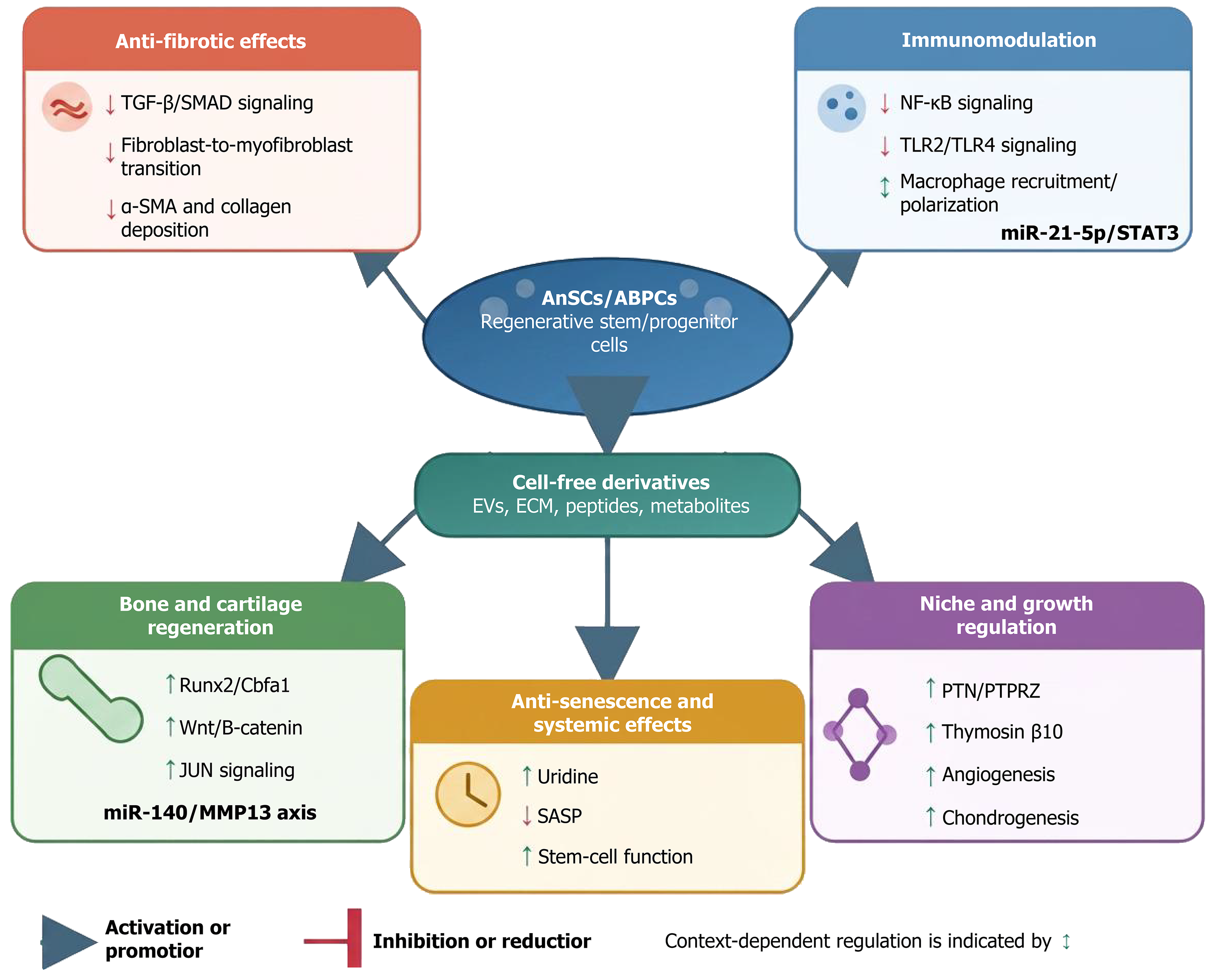
Figure 3 Simplified proposed model of signaling pathways associated with antler stem cell- and derivative-mediated tissue repair.
Antler stem cells (AnSCs), antler blastema progenitor cells (ABPCs), and their cell-free derivatives regulate tissue repair through interconnected anti-fibrotic, immunomodulatory, osteochondral, angiogenic, and anti-senescence pathways. Suppression of transforming growth factor-β/SMAD signaling and the fibroblast-to-myofibroblast transition reduces alpha-smooth muscle actin expression and collagen deposition. Nuclear factor kappa B and Toll-like receptor 2 (TLR2)/TLR4 inhibition, together with context-dependent regulation of macrophage recruitment and polarization, mediates immunomodulation. Runx2/Cbfa1, Wnt/β-catenin, JUN, and the miR-140/matrix metalloproteinase-13 axis regulate bone and cartilage regeneration. PTN/PTPRZ and thymosin β10 contribute to niche regulation, angiogenesis, and chondrogenesis, whereas the miR-21-5p/signal transducer and activator of transcription 3 axis and uridine are associated with wound healing and anti-senescence activity, respectively. TGF: Transforming growth factor; α-SMA: Alpha-smooth muscle actin; AnSCs: Antler stem cells; ABPCs: Antler blastema progenitor cells; NF-κB: Nuclear factor kappa B; TLR: Toll-like receptor; STAT3: Signal transducer and activator of transcription 3; EVs: Extracellular vesicles; ECM: Extracellular matrix; MMP13: Matrix metalloproteinase-13; SASP: Senescence-associated secretory phenotype.
- Citation: He Y, Li JP, Ma C, Zhang GK, Li CY. Potential therapeutic applications of antler stem cells and their derivatives. World J Stem Cells 2026; 18(10): 125616
- URL: https://www.wjgnet.com/1948-0210/full/v18/i10/125616.htm
- DOI: https://dx.doi.org/10.4252/wjsc.125616