
















Copyright: ©Author(s) 2026.
World J Stem Cells. Oct 26, 2026; 18(10): 125616
Published online Oct 26, 2026. doi: 10.4252/wjsc.125616
Published online Oct 26, 2026. doi: 10.4252/wjsc.125616
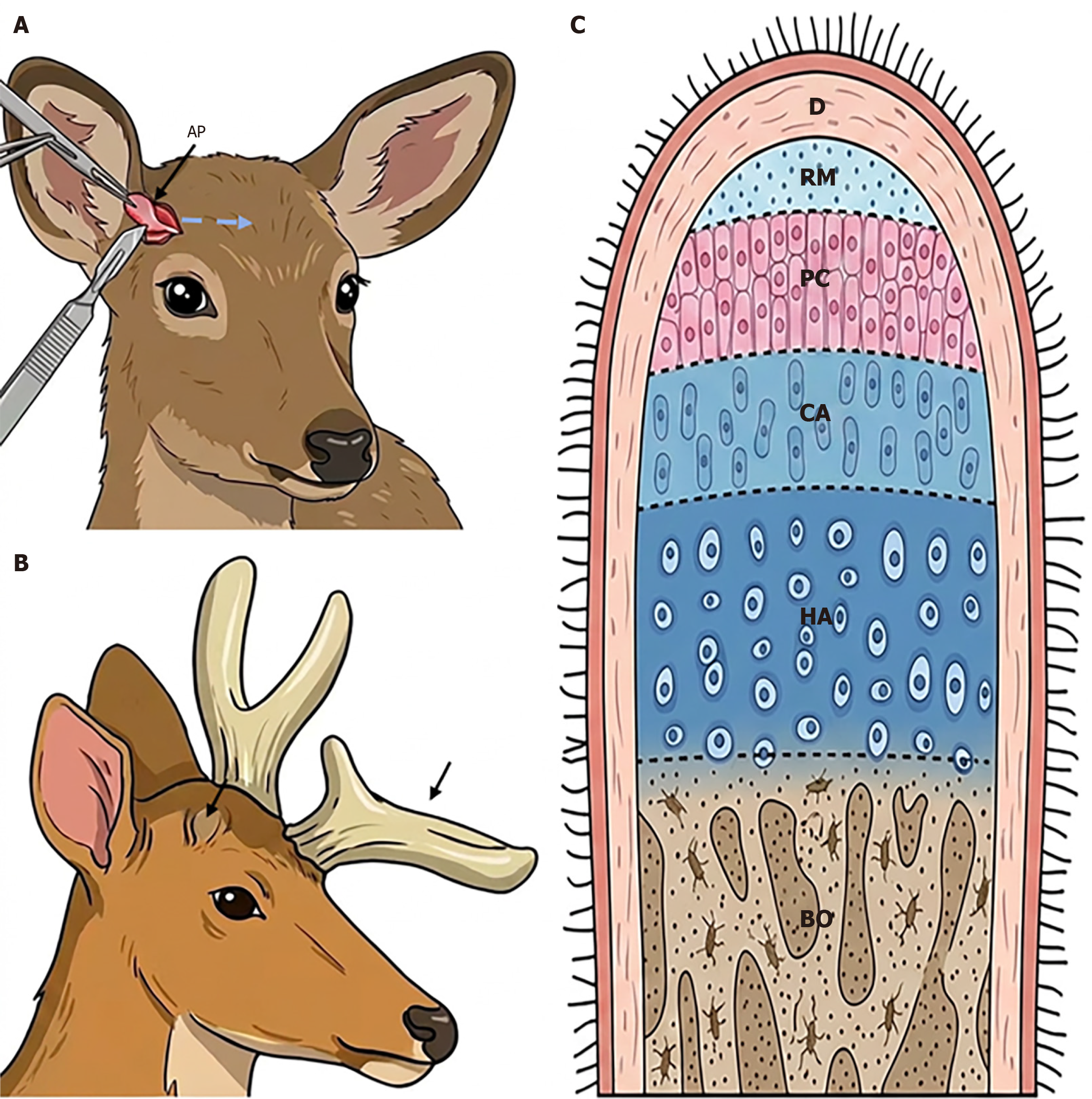
Figure 2 Antlerogenic periosteum deletion and transplantation, and histological structure of a growing antler tip.
A: Antlerogenic periosteum (AP) deletion (right side). Note that the deleted AP will be transplanted autologously onto the forehead region; B: Outcomes of AP deletion and transplantation. Note that the AP-less crest region failed to develop a pedicle and antler (arrow), but the transplanted forehead region initiated ectopic antler formation (arrowhead); C: Histological structure of a growing antler tip. Note that the reserve mesenchyme layer is located in the outermost position and immediately under the dermis of velvet skin. AP: Antlerogenic periosteum; D: Dermis; RM: Reserve mesenchyme; PC: Pre-cartilage; CA: Cartilage; HA: Hypertrophic cartilage; BO: Bone.
- Citation: He Y, Li JP, Ma C, Zhang GK, Li CY. Potential therapeutic applications of antler stem cells and their derivatives. World J Stem Cells 2026; 18(10): 125616
- URL: https://www.wjgnet.com/1948-0210/full/v18/i10/125616.htm
- DOI: https://dx.doi.org/10.4252/wjsc.125616