
















Copyright: ©Author(s) 2026.
World J Stem Cells. Oct 26, 2026; 18(10): 124640
Published online Oct 26, 2026. doi: 10.4252/wjsc.124640
Published online Oct 26, 2026. doi: 10.4252/wjsc.124640
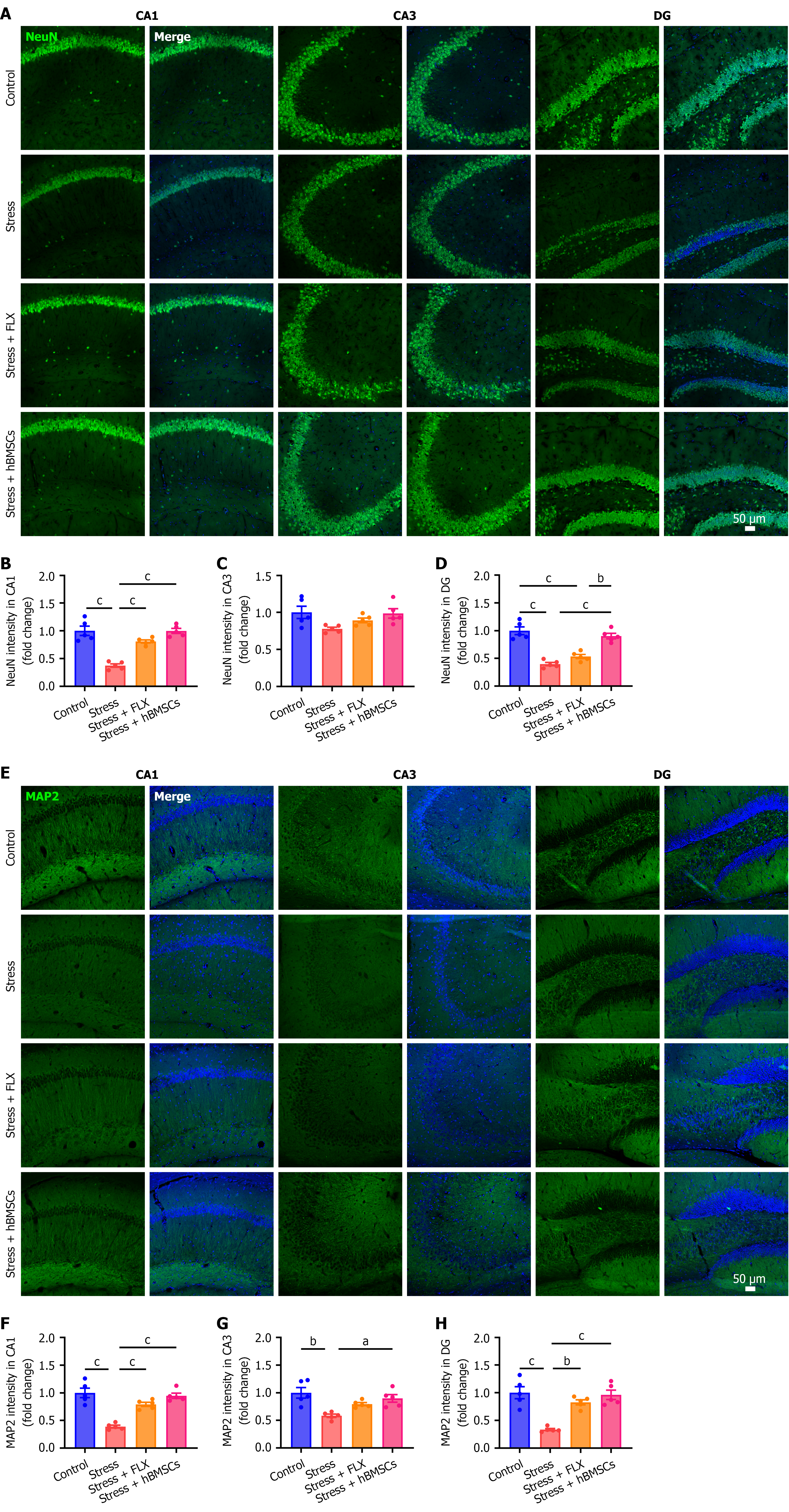
Figure 7 Human bone-marrow-derived mesenchymal stem cells exerted neuroprotective effects on the hippocampus of stressed mice.
A: Representative immunofluorescence images of NeuN in the different subregions of the hippocampus; B-D: Quantification of relative fluorescence intensity of NeuN in CA1 (B), CA3 (C), and dentate gyrus (DG) (D) regions; E: Representative immunofluorescence images of MAP2 in the different subregions of the hippocampus; F-H: Quantification of relative fluorescence intensity of MAP2 in CA1 (F), CA3 (G), and DG (H) regions. aP < 0.05, bP < 0.01, cP < 0.001. Data were analyzed using one-way analysis of variance followed by Tukey’s multiple-comparisons test (mean ± SEM, n = 5 per group). FLX: Fluoxetine; hBMSCs: Human bone-marrow-derived mesenchymal stem cells; DG: Dentate gyrus.
- Citation: Liu Y, Wu JF, Xu MY, Kang J, Ye AH, Liu B, Liu KZ. Human bone marrow-derived mesenchymal stem cells alleviate fluoxetine-resistant depression-like behaviors in mice by multi-target effects in the hippocampus. World J Stem Cells 2026; 18(10): 124640
- URL: https://www.wjgnet.com/1948-0210/full/v18/i10/124640.htm
- DOI: https://dx.doi.org/10.4252/wjsc.124640