
















Copyright: ©Author(s) 2026.
World J Stem Cells. Oct 26, 2026; 18(10): 119325
Published online Oct 26, 2026. doi: 10.4252/wjsc.119325
Published online Oct 26, 2026. doi: 10.4252/wjsc.119325
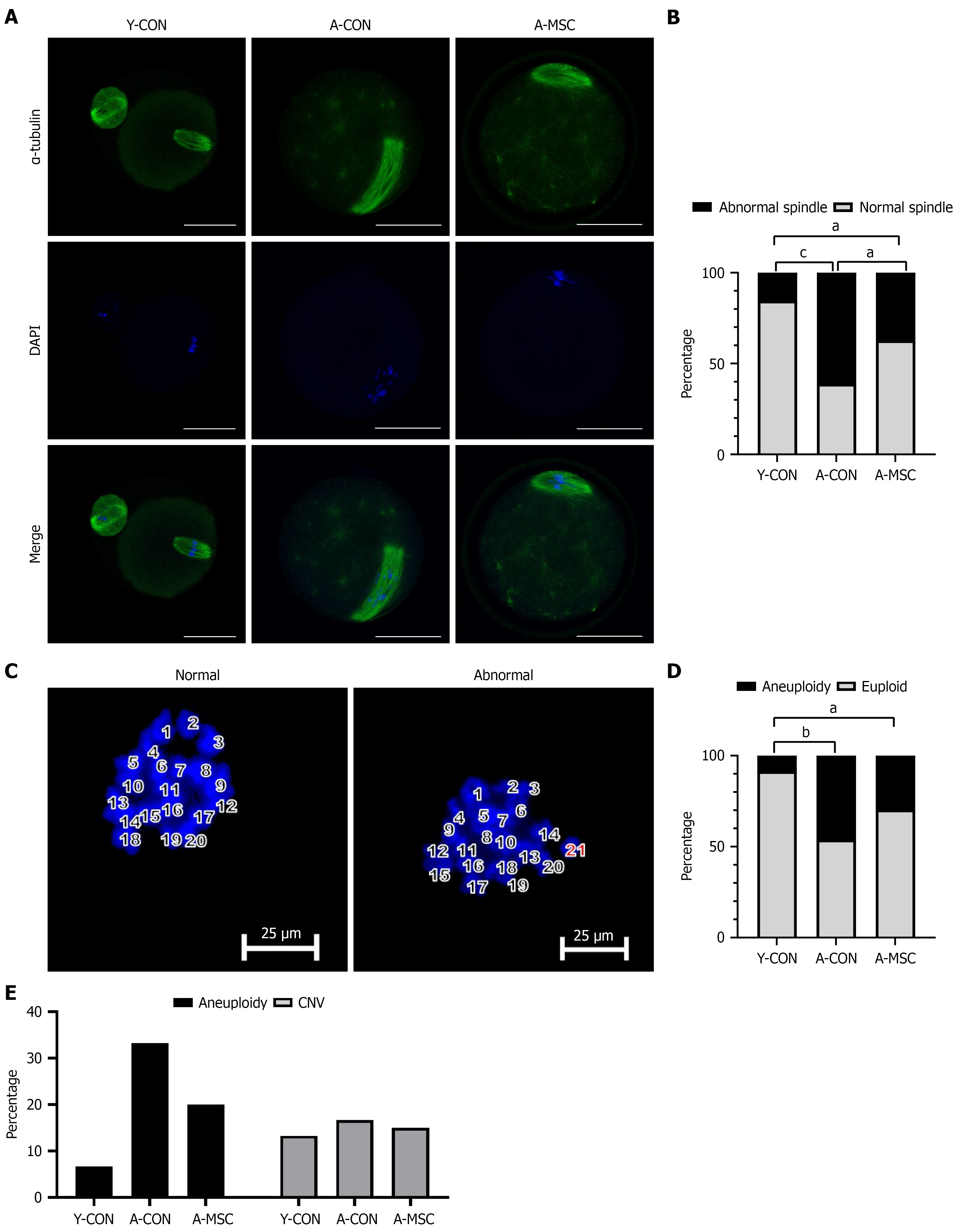
Figure 4 Umbilical cord-derived mesenchymal stem cell transplantation improves the meiotic apparatus and reduces aneuploidy tendency in oocytes from aged mice.
A: Representative immunofluorescence images of meiotic spindles (green, α-tubulin) and chromosomes (blue, DAPI) in metaphase II (MII) oocytes. Scale bar = 20 μm; B: Quantitative analysis of the percentage of oocytes with normal spindle morphology. Umbilical cord-derived mesenchymal stem cell transplantation (A-MSC) significantly rescued the age-related decline in normal spindle formation; C: Representative images of chromosome spreads from MII oocytes, showing a normal euploid count (20 chromosomes) and an abnormal aneuploid count (21 chromosomes). Scale bar = 20 μm; D: Quantitative analysis of the percentage of oocytes with a normal chromosome count (20); E: Bar chart showing the percentages of aneuploid oocytes and oocytes with copy number variations across the different groups. Data are presented as mean ± SEM. aP < 0.05, bP < 0.01, cP < 0.001. Y-CON: Young control; A-CON: Aged control; A-MSC: Umbilical cord-derived mesenchymal stem cell transplantation; CNV: Copy number variation.
- Citation: Zhang JN, Tian S, Guo XM, Zhang Q, Shang J, Yao YQ. Therapeutic potential of umbilical cord-derived mesenchymal stem cells in ameliorating ovarian aging: Insights from single-cell analysis. World J Stem Cells 2026; 18(10): 119325
- URL: https://www.wjgnet.com/1948-0210/full/v18/i10/119325.htm
- DOI: https://dx.doi.org/10.4252/wjsc.119325