
















Copyright: ©Author(s) 2026.
World J Stem Cells. Oct 26, 2026; 18(10): 119325
Published online Oct 26, 2026. doi: 10.4252/wjsc.119325
Published online Oct 26, 2026. doi: 10.4252/wjsc.119325
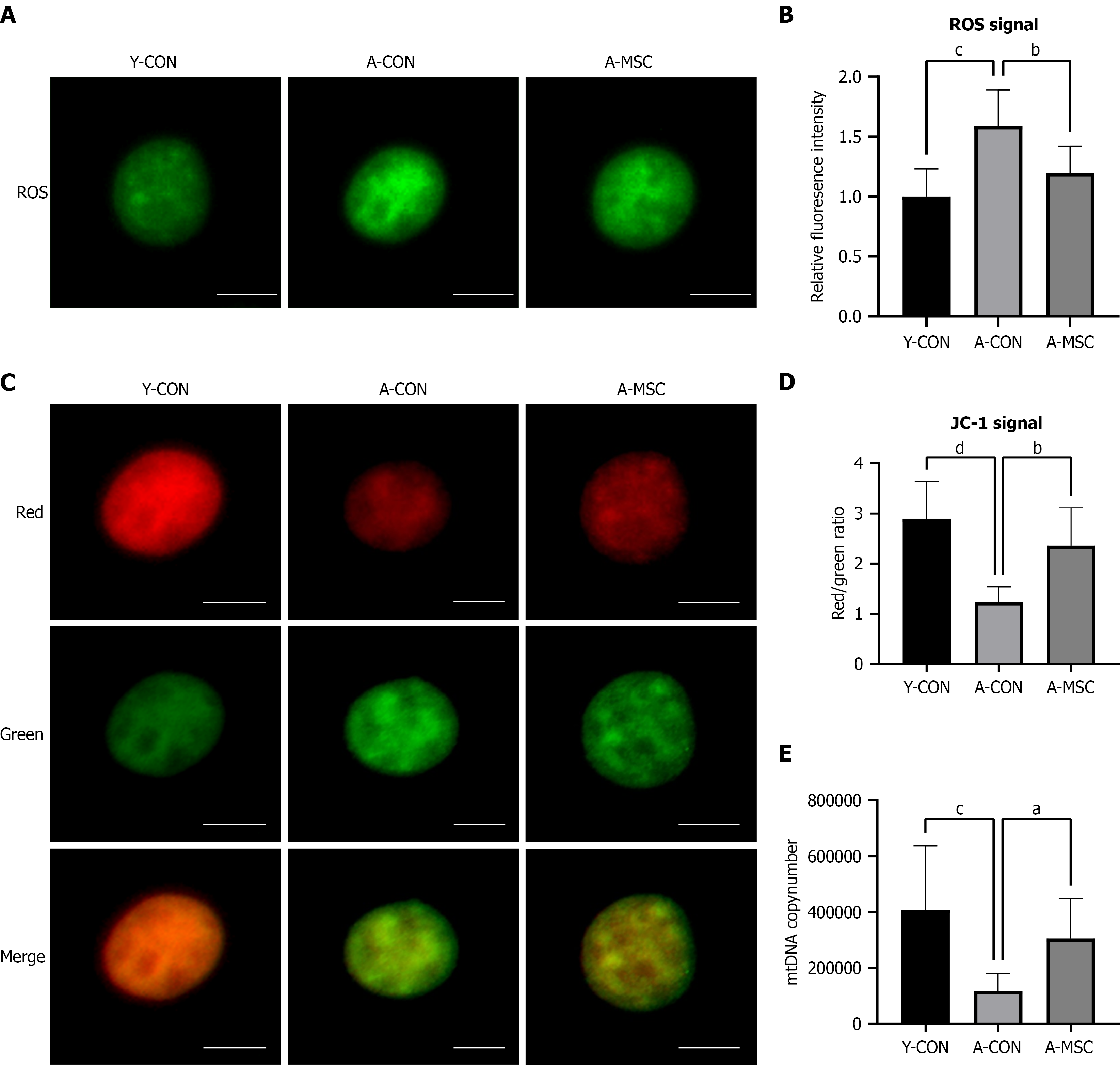
Figure 3 Umbilical cord-derived mesenchymal stem cell transplantation ameliorates oocyte mitochondrial function in aged mice.
A: Representative images of reactive oxygen species (ROS) levels in oocytes, detected by DCFH-DA fluorescence; B: Quantitative analysis of ROS levels in oocytes. ROS levels were significantly elevated in the aged control (A-CON) group and were reduced following umbilical cord-derived mesenchymal stem cell transplantation (A-MSC); C: Assessment of mitochondrial membrane potential (ΔΨm) using JC-1 staining. Representative fluorescence images are shown (red: J-aggregates, high ΔΨm; green: J-monomers, low ΔΨm); D: Quantitative analysis of the JC-1 red-to-green fluorescence intensity ratio. The reduced ratio observed in A-CON oocytes was significantly restored by A-MSC; E: Absolute quantification of mitochondrial DNA (mtDNA) copy number in individual metaphase II oocytes. The marked depletion of mtDNA copy number in the A-CON group was significantly rescued by A-MSC. Scale bar = 20 μm. Data are presented as mean ± SEM. aP < 0.05, bP < 0.01, cP < 0.001, dP < 0.0001. Y-CON: Young control; A-CON: Aged control; A-MSC: Umbilical cord-derived mesenchymal stem cell transplantation; ROS: Reactive oxygen species; mtDNA: Mitochondrial DNA.
- Citation: Zhang JN, Tian S, Guo XM, Zhang Q, Shang J, Yao YQ. Therapeutic potential of umbilical cord-derived mesenchymal stem cells in ameliorating ovarian aging: Insights from single-cell analysis. World J Stem Cells 2026; 18(10): 119325
- URL: https://www.wjgnet.com/1948-0210/full/v18/i10/119325.htm
- DOI: https://dx.doi.org/10.4252/wjsc.119325