










BPG is committed to discovery and dissemination of knowledge
Basic Study
Copyright: ©Author(s) 2026.
World J Stem Cells. Oct 26, 2026; 18(10): 119325
Published online Oct 26, 2026. doi: 10.4252/wjsc.119325
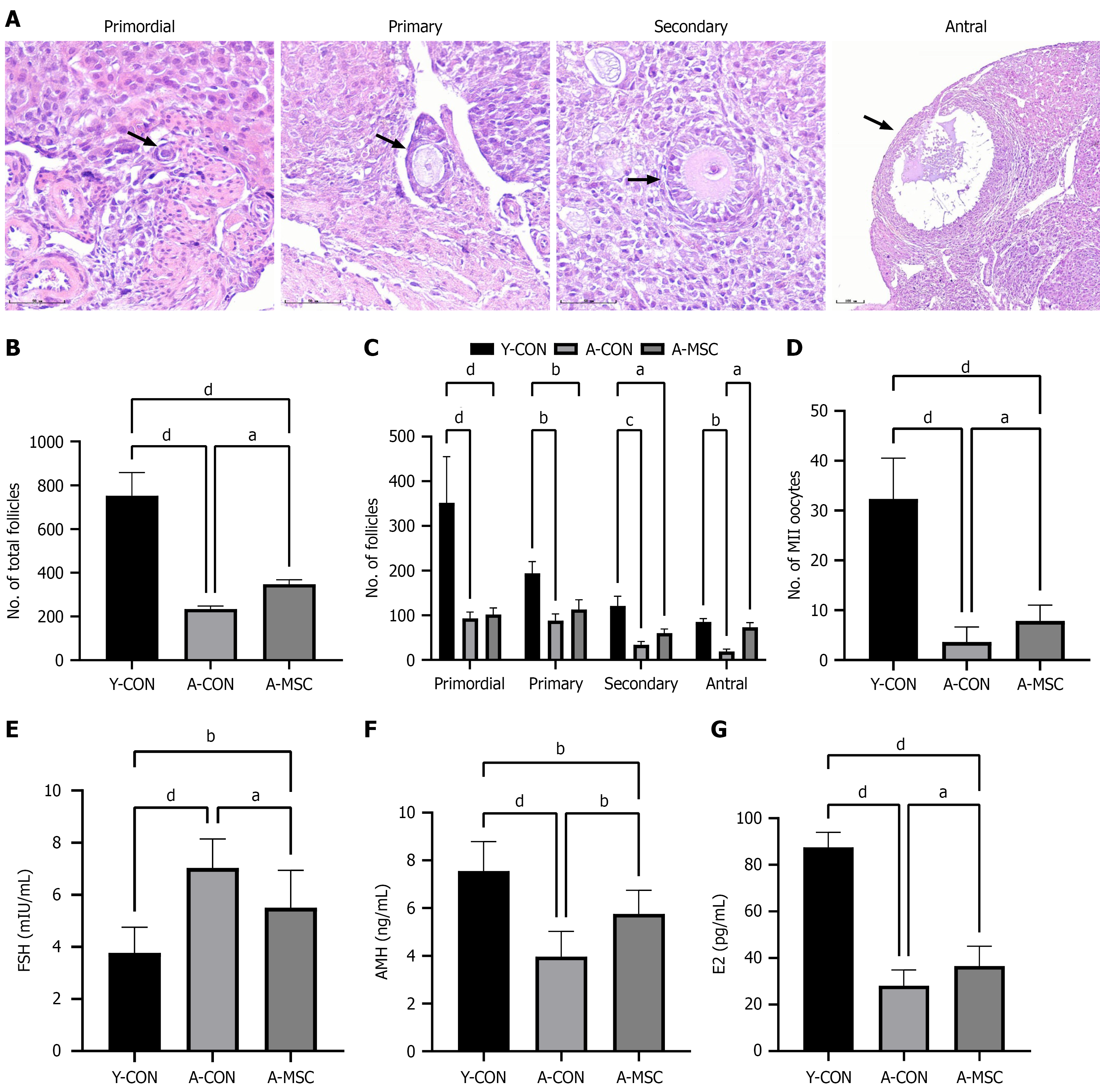
Figure 2
Figure 2 Umbilical cord-derived mesenchymal stem cell transplantation enhances ovarian reserve and function in aged mice. A: Representative hematoxylin and eosin-stained images of ovarian sections showing different follicular developmental stages (arrows). Primordial follicle: An oocyte surrounded by a single layer of flattened granulosa cells, mainly located in the ovarian cortex. Primary follicle: An enlarging oocyte enclosed by a single layer of cuboidal granulosa cells. Secondary follicle: An oocyte surrounded by multiple layers of granulosa cells. Antral follicle: An oocyte surrounded by multiple layers of granulosa cells, with visible antral cavity formation. Scale bar = 100 μm; B: Number of follicles at each developmental stage per ovary; C: Total follicle count per ovary. Umbilical cord-derived mesenchymal stem cell transplantation (A-MSC) significantly increased the total follicle count, with a significant increase in the number of antral follicles; D: Number of metaphase II (MII) oocytes collected after superovulation. The A-MSC group yielded significantly more MII oocytes than the aged control group. Serum hormone levels measured by enzyme-linked immunosorbent assay; E: Follicle-stimulating hormone (FSH) level; F: Anti-Müllerian hormone (AMH) level; G: Estradiol (E2) level. A-MSC significantly reduced FSH levels and increased AMH and E2 levels in aged mice. Data are presented as mean ± SEM. aP < 0.05, bP < 0.01, cP < 0.001, and dP < 0.0001. Y-CON: Young control; A-CON: Aged control; A-MSC: Umbilical cord-derived mesenchymal stem cell transplantation; FSH: Follicle-stimulating hormone; AMH: Anti-Müllerian hormone; E2: Estradiol.


Write to the Help Desk