
















©The Author(s) 2026.
World J Stem Cells. Jan 26, 2026; 18(1): 113614
Published online Jan 26, 2026. doi: 10.4252/wjsc.v18.i1.113614
Published online Jan 26, 2026. doi: 10.4252/wjsc.v18.i1.113614
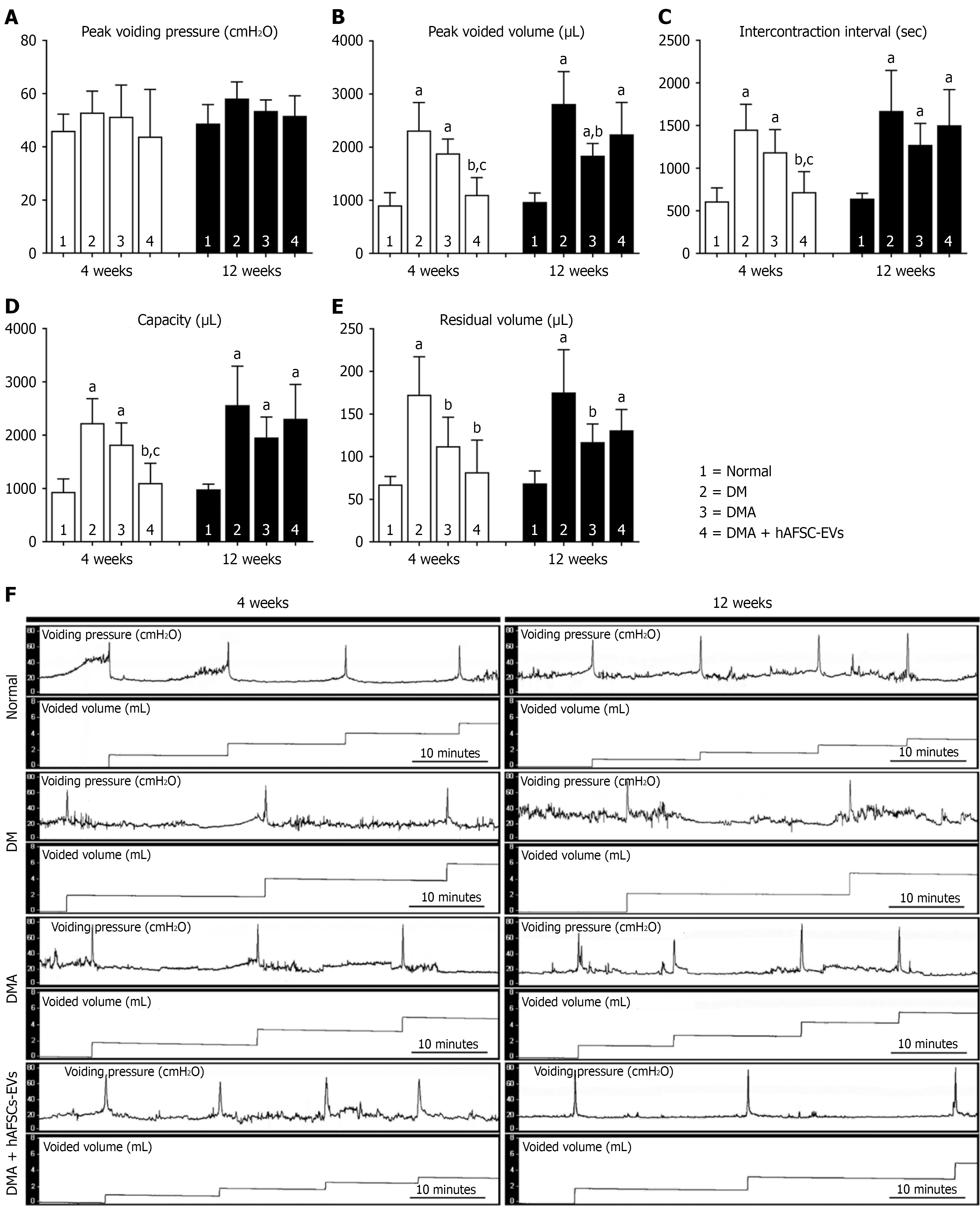
Figure 3 Cystometric evaluation of bladder function.
A-F: At 4 weeks and 12 weeks after induction, peak voided volume, intercontraction interval and bladder capacity were significantly increased in both diabetes mellitus (DM) and diabetic atherosclerosis (DMA) groups compared with the control group, whereas residual volume was elevated only in the DM group. Furthermore, compared with the DM group, DMA rats had significantly reduced residual volume at 4 weeks and 12 weeks after induction, and peak voided volume was decreased at 12 weeks. In DMA rats treated with human amniotic fluid stem cell-derived extracellular vesicles, peak voided volume, intercontraction interval and bladder capacity were significantly reduced at 4 weeks after induction. P values are shown in each comparison. Cystometric variables include: Peak voiding pressure (A); peak voided volume (B); intercontraction interval (C); bladder capacity (D); residual volume (E); representative cystometry tracings for normal, DM, DMA, and DMA + human amniotic fluid stem cell-derived extracellular vesicles groups at 4 weeks and 12 weeks after induction (F). aP < 0.05 vs normal control, bP < 0.05 vs diabetes mellitus, cP < 0.05 vs diabetic atherosclerosis. DM: Diabetes mellitus; DMA: Diabetic atherosclerosis; hAFSC-EVs: Human amniotic fluid stem cell-derived extracellular vesicles.
- Citation: Liang CC, Lin YH, Liang CY, Ro A, Huang YH, Shaw SW. Extracellular vesicles derived from human amniotic fluid stem cells improve bladder dysfunction in rat model of diabetic atherosclerosis. World J Stem Cells 2026; 18(1): 113614
- URL: https://www.wjgnet.com/1948-0210/full/v18/i1/113614.htm
- DOI: https://dx.doi.org/10.4252/wjsc.v18.i1.113614