
















©The Author(s) 2026.
World J Stem Cells. Jan 26, 2026; 18(1): 113614
Published online Jan 26, 2026. doi: 10.4252/wjsc.v18.i1.113614
Published online Jan 26, 2026. doi: 10.4252/wjsc.v18.i1.113614
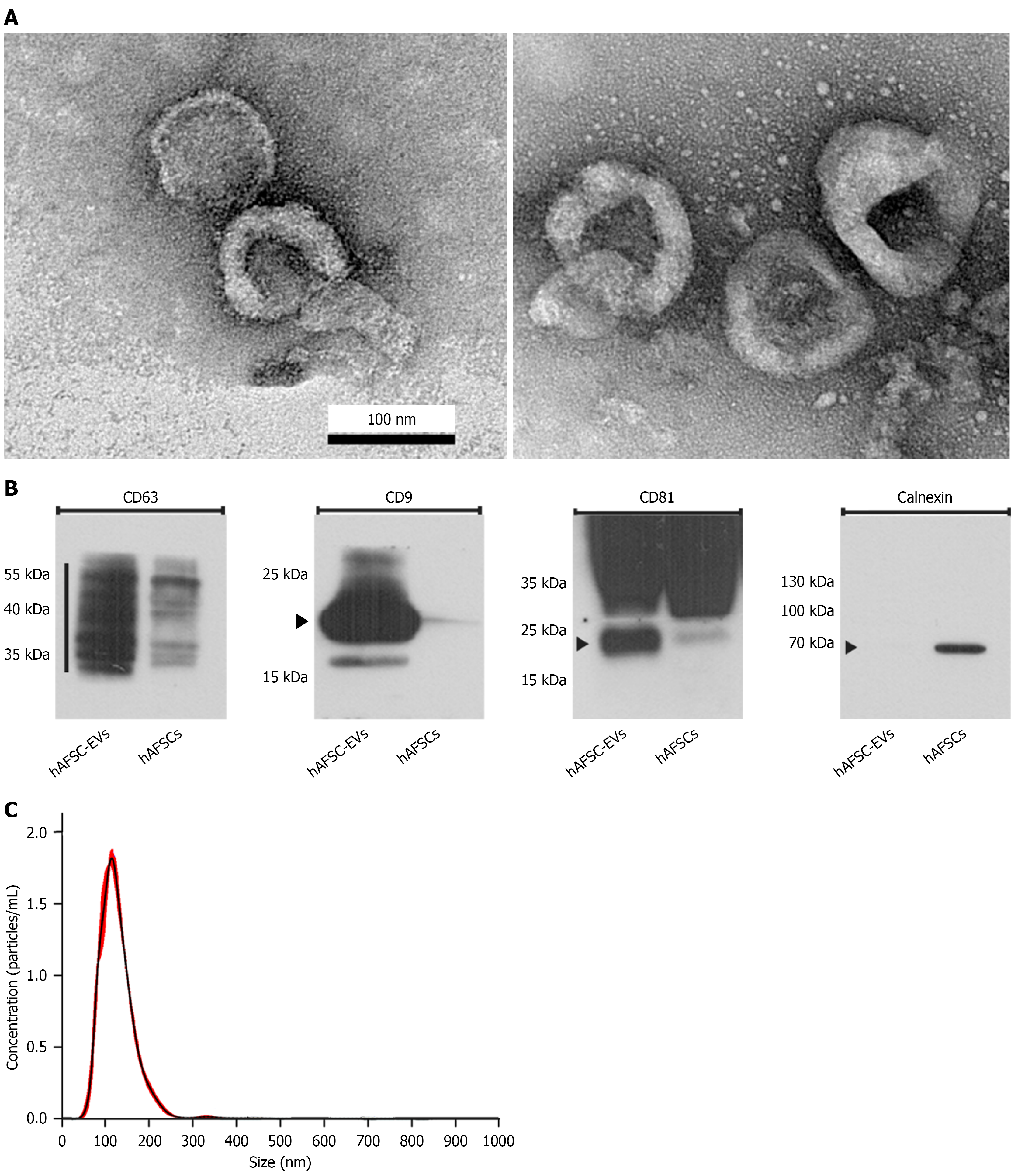
Figure 2 Characterization of human amniotic fluid stem cell-derived extracellular vesicles.
Transmission electron microscopy shows that human amniotic fluid stem cell-derived extracellular vesicles exhibit a bilayered spherical morphology with diameters ranging from 50 nm to 150 nm. Bars indicate 100 nm. Western blot analysis confirms the expression of exosomal surface markers CD63, CD81, and CD9. Nanoparticle tracking analysis reveals that the majority of human amniotic fluid stem cell-derived extracellular vesicles fall within the 50-150 nm size range. A: Transmission electron microscopy; B: Western blot analysis; C: Nanoparticle tracking analysis. hAFSC-EVs: Human amniotic fluid stem cell-derived extracellular vesicles; hAFSCs: Human amniotic fluid stem cells.
- Citation: Liang CC, Lin YH, Liang CY, Ro A, Huang YH, Shaw SW. Extracellular vesicles derived from human amniotic fluid stem cells improve bladder dysfunction in rat model of diabetic atherosclerosis. World J Stem Cells 2026; 18(1): 113614
- URL: https://www.wjgnet.com/1948-0210/full/v18/i1/113614.htm
- DOI: https://dx.doi.org/10.4252/wjsc.v18.i1.113614