











BPG is committed to discovery and dissemination of knowledge
Basic Study
©The Author(s) 2026.
World J Stem Cells. Jan 26, 2026; 18(1): 112278
Published online Jan 26, 2026. doi: 10.4252/wjsc.v18.i1.112278
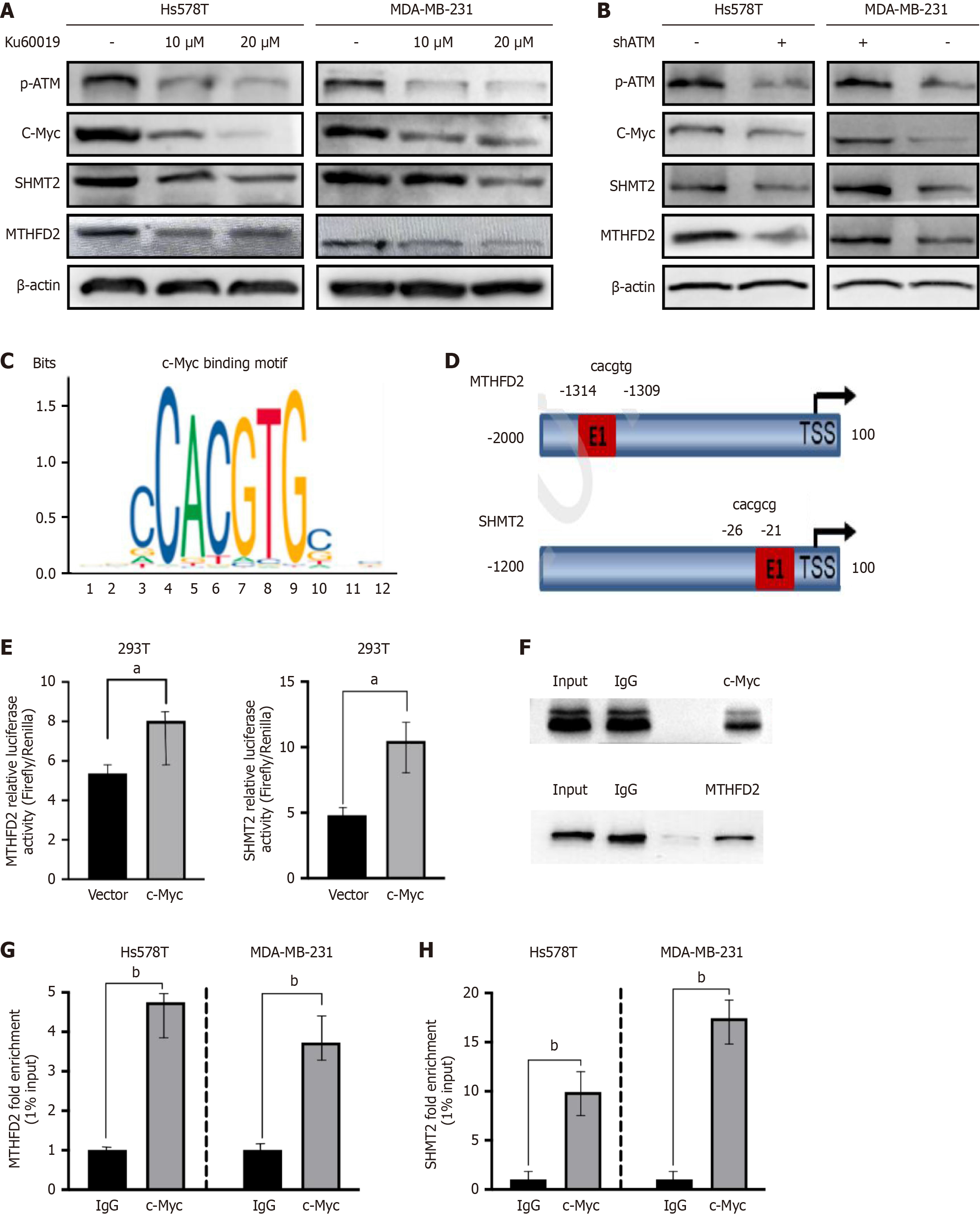
Figure 4
Figure 4 Oxidized ataxia telangiectasia mutated promotes serine hydroxymethyltransferase 2 and methylenetetrahydrofolate dehydrogenase 2 expression through c-Myc. A and B: Western blot analysis of phosphorylated ataxia telangiectasia mutated, c-Myc, serine hydroxymethyltransferase 2 (SHMT2), and methylenetetrahydrofolate dehydrogenase 2 (MTHFD2) in Hs578T and MDA-MB-231 cells after treatment with Ku60019 and ataxia telangiectasia mutated knockdown; C: Consensus c-Myc binding motif; D: Schematic representation of predicted c-Myc binding sites in the human MTHFD2 and SHMT2 promoter regions; E: Luciferase assay showed SHMT2 and MTHFD2 relative luciferase activity; F: Representative chromatin immunoprecipitation (ChIP)-polymerase chain reaction (PCR) showing c-Myc occupancy at the MTHFD2 and SHMT2 promoters; input and immunoglobulin G served as controls; G and H: ChIP-quantitative PCR analysis demonstrating c-Myc enrichment at the MTHFD2 (G) and SHMT2 (H) promoters in Hs578T and MDA-MB-231 cells. ChIP-quantitative PCR enrichment expressed as % input relative to immunoglobulin G. Data were presented as mean ± SD (n = 3). aP < 0.05; bP < 0.01. p-ATM: Phosphorylated ataxia telangiectasia mutated; MTHFD2: Methylenetetrahydrofolate dehydrogenase 2; SHMT2: Serine hydroxymethyltransferase 2; IgG: Immunoglobulin G.


Write to the Help Desk