
















©The Author(s) 2026.
World J Stem Cells. Jan 26, 2026; 18(1): 112278
Published online Jan 26, 2026. doi: 10.4252/wjsc.v18.i1.112278
Published online Jan 26, 2026. doi: 10.4252/wjsc.v18.i1.112278
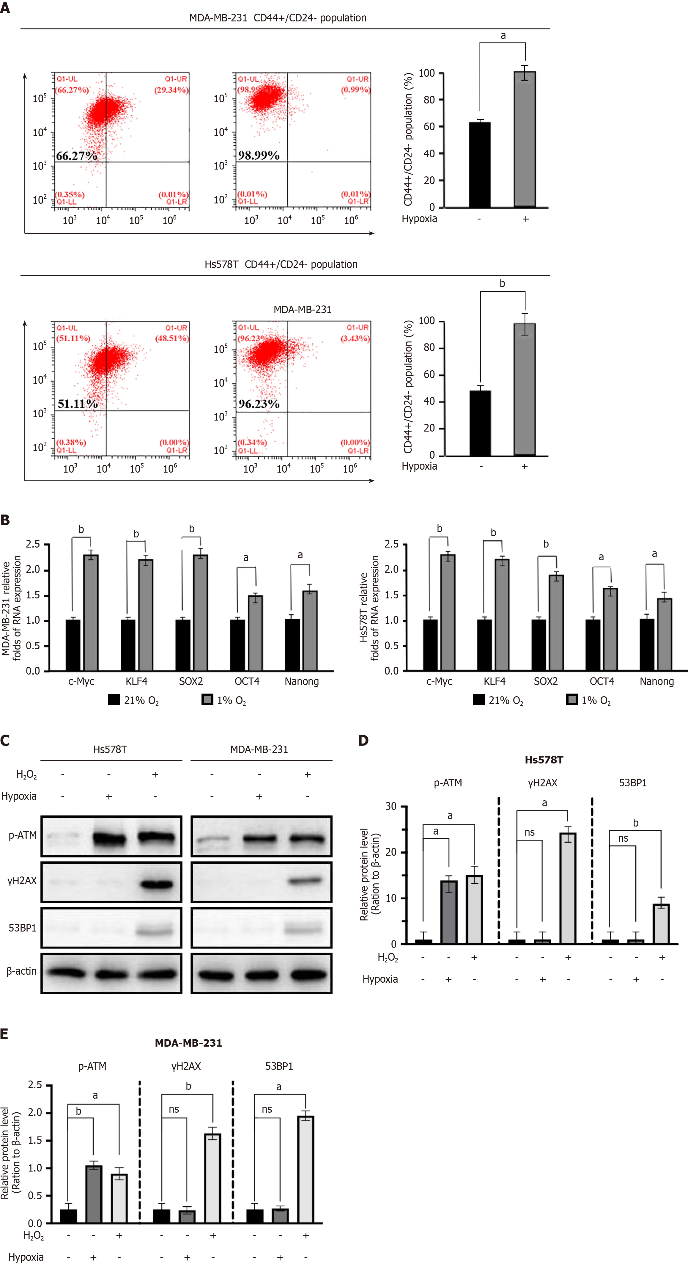
Figure 1 Hypoxia promotes enrichment and DNA damage-independent oxidative activation of ataxia telangiectasia mutated.
A: Flow cytometry analysis of CD44+/CD24- cell populations in Hs578T and MDA-MB-231 cells after exposure to hypoxia or normoxia; B: Quantitative reverse transcriptase polymerase chain reaction analysis of cancer stem cells-associated genes (c-Myc, octamer-binding protein 4, Kruppel-like factor 4, sex-determining region Y-box 2, NANOG) in mammosphere cultures under hypoxia; C-E: Western blot analysis of phosphorylated ataxia telangiectasia mutated, γH2AX, and 53BP1 in Hs578T and MDA-MB-231 cancer stem cells under normoxia, hypoxia, or H2O2 treatment. Data were presented as mean ± SD (n = 3). aP < 0.05; bP < 0.01; ns: not significant. KLF4: Kruppel-like factor 4; SOX2: Sex-determining region Y-box 2; OCT4: Octamer-binding protein 4; p-ATM: Phosphorylated ataxia telangiectasia mutated.
- Citation: Yang D, Ou YL, Wang SH, Jin HJ, Chen SH, Han R, Zhang H. Hypoxia facilitates triple-negative breast cancer stem cells enrichment and stemness maintenance through oxidized ataxia telangiectasia mutated-induced one-carbon metabolism. World J Stem Cells 2026; 18(1): 112278
- URL: https://www.wjgnet.com/1948-0210/full/v18/i1/112278.htm
- DOI: https://dx.doi.org/10.4252/wjsc.v18.i1.112278