
















©The Author(s) 2025.
World J Stem Cells. Sep 26, 2025; 17(9): 109715
Published online Sep 26, 2025. doi: 10.4252/wjsc.v17.i9.109715
Published online Sep 26, 2025. doi: 10.4252/wjsc.v17.i9.109715
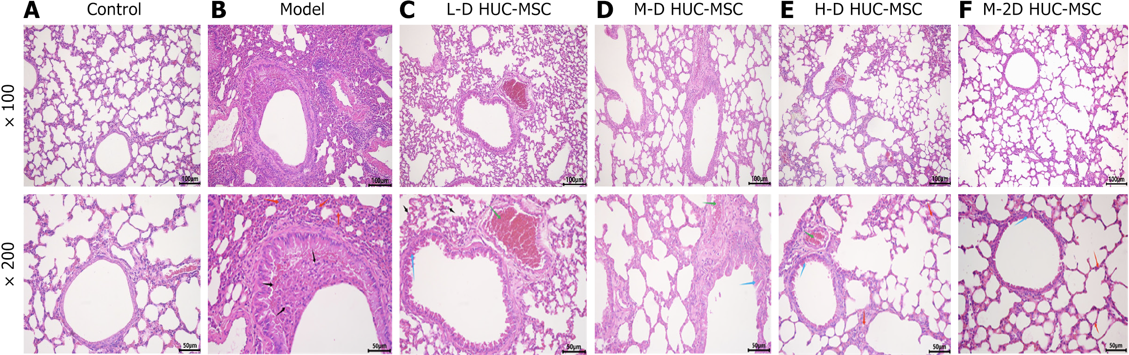
Figure 5 Pathological features of human umbilical cord blood-derived mesenchymal stem cell in attenuating airway inflammation in ovalbumin-induced asthmatic mice.
A-F: Hematoxylin and eosin staining images of control, model, low dosage, medium dosage, high dosage, and two medium dosage human umbilical cord blood-derived mesenchymal stem cell treated lung tissue of mice. Hematoxylin and eosin staining showed that the control group displayed normal lung tissue architecture and cellular morphology, with no inflammatory cell infiltration. Conversely, the model group demonstrated pronounced inflammatory cell infiltration (indicated by black arrows), thickening of the alveolar walls (indicated by red arrows), sparse inflammatory cells (black arrows), desquamation of bronchial epithelial cells (indicated by blue arrows), and vascular congestion (indicated by green arrows) across the various dose groups. Notably, the group receiving a double medium dose of human umbilical cord blood-derived mesenchymal stem cell exhibited significant improvement in the condition of the alveolar walls compared to the model group (red arrows). L-D HUC-MSC: Low dosage (1 × 10⁵) of human umbilical cord blood-derived mesenchymal stem cell; M-D HUC-MSC: Medium dosage (2 × 10⁵) of human umbilical cord blood-derived mesenchymal stem cell; H-D HUC-MSC: High dosage (4 × 10⁵) of human umbilical cord blood-derived mesenchymal stem cell; M-2D HUC-MSC: Medium dosage (2 × 10⁵) of human umbilical cord blood-derived mesenchymal stem cell twice.
- Citation: Chen QH, Zheng JY, Zhu YQ, Zhang JY, Lin CY, Zhuang XE, Cheng J, Huang XY. Exploring the critical therapeutic window: Dose-frequency optimization of human umbilical cord mesenchymal stem cells for preclinical asthma treatment. World J Stem Cells 2025; 17(9): 109715
- URL: https://www.wjgnet.com/1948-0210/full/v17/i9/109715.htm
- DOI: https://dx.doi.org/10.4252/wjsc.v17.i9.109715