
















©The Author(s) 2025.
World J Stem Cells. Sep 26, 2025; 17(9): 108049
Published online Sep 26, 2025. doi: 10.4252/wjsc.v17.i9.108049
Published online Sep 26, 2025. doi: 10.4252/wjsc.v17.i9.108049
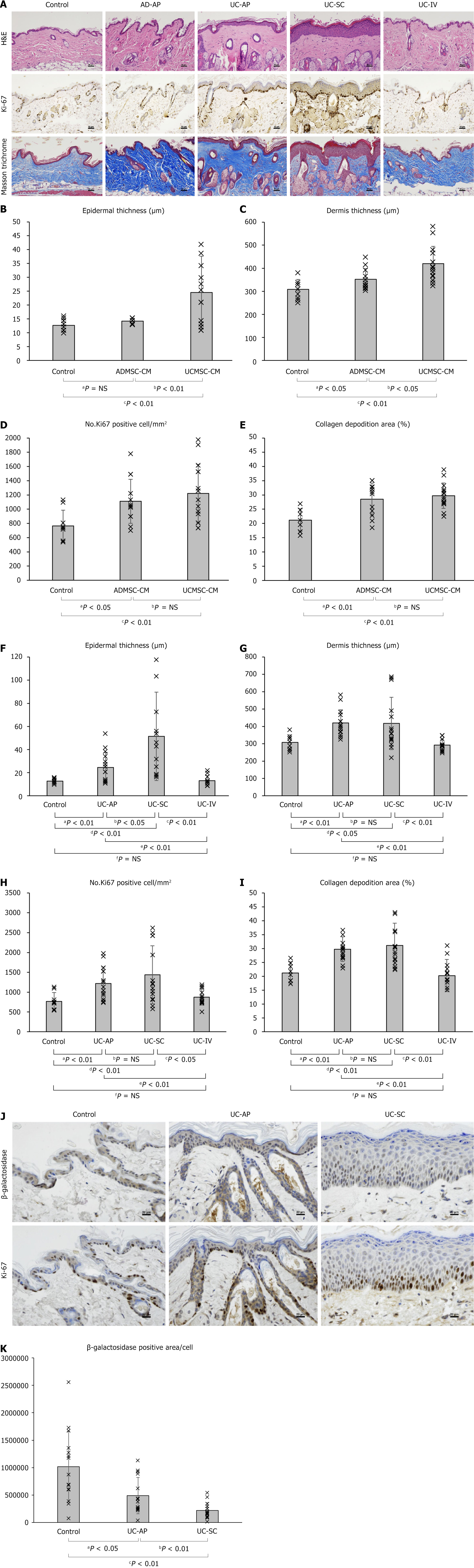
Figure 4 In vivo effectiveness test of umbilical cord-derived mesenchymal stem cell-conditioned medium.
A: Representative histological images of aged mouse skin stained with hematoxylin-eosin, Ki-67, and Masson’s Trichrome stains. Scale bar = 50 μm; B-E: Quantitative analysis of epidermal thickness (B), dermal thickness (C), number of Ki-67-positive cells per unit area (D), and collagen deposition area (E) according to the type of reagent; F-I: Quantitative analysis of epidermal thickness (F), dermal thickness (G) number of Ki-67-positive cells per unit area (H), and collagen deposition area (I) according to the method used for umbilical cord-derived mesenchymal stem cell-conditioned medium administration; J: Serial sections of aged mouse skin stained with Ki-67 and β-galactosidase. Scale bar = 20 μm; K: Quantification of β-galactosidase expression per epidermal cell. All quantitative data (B-I and K) were obtained from color-based image analysis of stained tissue sections. NS: Not significant; AD-AP: Adipose-derived mesenchymal stem cell-conditioned medium application; UC-AP: Umbilical cord-derived mesenchymal stem cell-conditioned medium application; UC-SC: Umbilical cord-derived mesenchymal stem cell-conditioned medium subcutaneous injection; UC-IV: Umbilical cord-derived mesenchymal stem cell-conditioned medium tail-vein injection; ADMSC-CM: Adipose-derived mesenchymal stem cell-conditioned medium; UCMSC-CM: Umbilical cord-derived mesenchymal stem cell-conditioned medium.
- Citation: Ahn H, Han HS, Lee KH. Efficacy and safety of umbilical cord-derived mesenchymal stem cell-conditioned media for preventing and treating skin aging. World J Stem Cells 2025; 17(9): 108049
- URL: https://www.wjgnet.com/1948-0210/full/v17/i9/108049.htm
- DOI: https://dx.doi.org/10.4252/wjsc.v17.i9.108049