Copyright
©The Author(s) 2025.
World J Stem Cells. Jun 26, 2025; 17(6): 106272
Published online Jun 26, 2025. doi: 10.4252/wjsc.v17.i6.106272
Published online Jun 26, 2025. doi: 10.4252/wjsc.v17.i6.106272
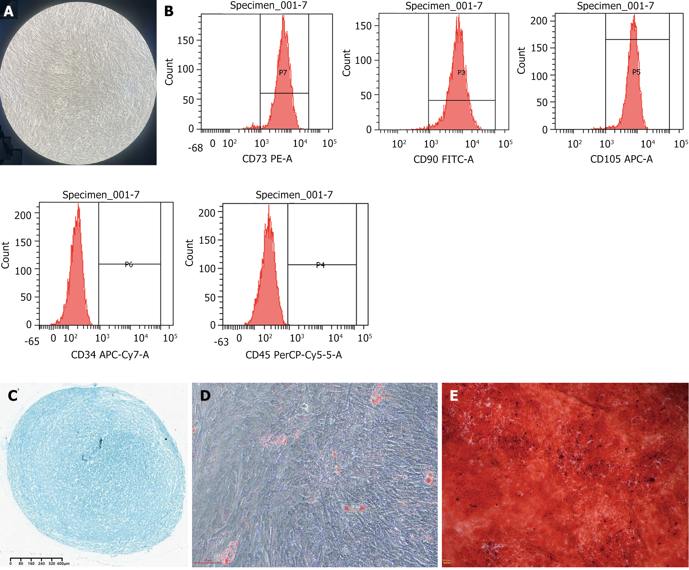
Figure 1 Isolation, culture, identification, and induced differentiation of human umbilical cord mesenchymal stem cells.
A: On day 15 of primary cell culture, human umbilical cord mesenchymal stem cells exhibited robust growth and over 90% confluence (magnification 100 ×); B: Identification of human umbilical cord mesenchymal stem cell surface antigens by flow cytometry; C: Induction of chondroblastic differentiation for three weeks resulted in the appearance of chondrocytes that stained blue with Alcian Blue (magnification 50 ×); D: Induction of adipogenic differentiation for two weeks resulted in the appearance of lipid droplets that stained red with Oil Red O (magnification 200 ×); E: Induction of osteogenic differentiation for three weeks led to the appearance of calcium nodules that stained red with Alizarin Red (magnification 100 ×).
- Citation: Zhang B, Gao BD, Su Y, Mi WJ, Zeng TX, Ma FF, Ha XQ. Human umbilical cord mesenchymal stem cells reduce platelet α-granule release in rats via the AKT/MEK/ERK pathway during acute exposure to high-altitude hypoxia. World J Stem Cells 2025; 17(6): 106272
- URL: https://www.wjgnet.com/1948-0210/full/v17/i6/106272.htm
- DOI: https://dx.doi.org/10.4252/wjsc.v17.i6.106272


