
















©The Author(s) 2025.
World J Stem Cells. Dec 26, 2025; 17(12): 112207
Published online Dec 26, 2025. doi: 10.4252/wjsc.v17.i12.112207
Published online Dec 26, 2025. doi: 10.4252/wjsc.v17.i12.112207
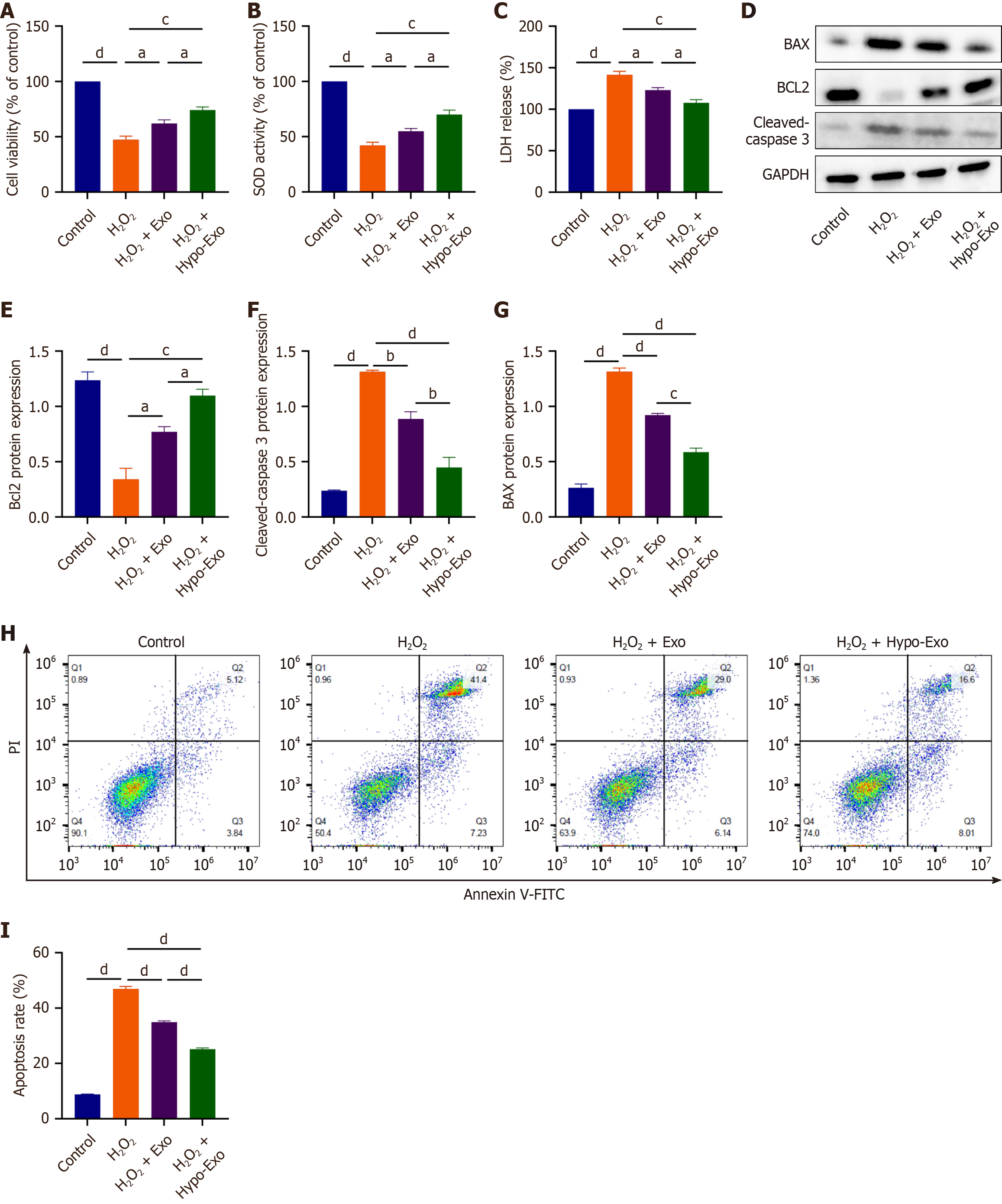
Figure 3 Human umbilical cord-derived mesenchymal stem cells exosomes ameliorated oxidative stress-induced apoptosis in H9C2 cells.
A: Different effects of normoxic counterparts (normo-Exos) and hypoxia-preconditioned mesenchymal stem cell-derived exosomes (hypo-Exos) on the viability of H9C2 cells; B: Different effects of normo-Exos and hypo-Exos on superoxide dismutase (SOD) activity in hydrogen peroxide (H2O2)-induced H9C2 cells; C: Different effects of normo-Exos and hypo-Exos on lactate dehydrogenase (LDH) release in H2O2-induced H9C2 cells; D: Expression of apoptosis-related protein was determined by western blotting; E-G: Quantitative analysis of the western blotting; H: Level of apoptosis was determined by flow cytometry; I: Statistical evaluation of flow cytometry. Data are presented as the mean ± standard error of the mean (n = 3). aP < 0.05, bP < 0.01, cP < 0.001, dP < 0.0001.
- Citation: Zeng ZF, Rao J, Xia XB, Chen XY, He HX, Liu B, Chen Q, Liu YD, Wang GJ, Cheng PC, Wang JN, Wang P, Yu Y, Wang ZN. Hypoxic preconditioned mesenchymal stem cell-derived exosomes alleviate oxidative stress-induced cardiomyocyte apoptosis through miR-486-5p. World J Stem Cells 2025; 17(12): 112207
- URL: https://www.wjgnet.com/1948-0210/full/v17/i12/112207.htm
- DOI: https://dx.doi.org/10.4252/wjsc.v17.i12.112207