
















©The Author(s) 2025.
World J Stem Cells. Dec 26, 2025; 17(12): 106128
Published online Dec 26, 2025. doi: 10.4252/wjsc.v17.i12.106128
Published online Dec 26, 2025. doi: 10.4252/wjsc.v17.i12.106128
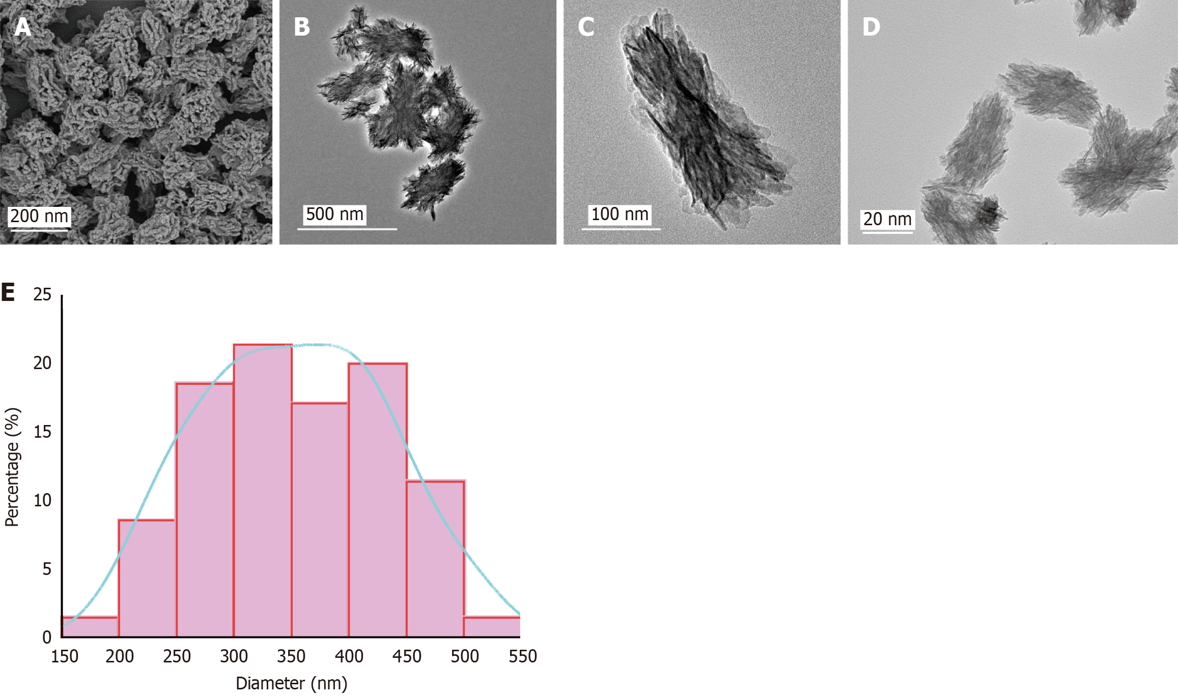
Figure 1 Ultrastructural morphology of calcifying nanoparticles observed by scanning electron microscope and transmission electron microscope.
A: Scanning electron microscope images of calcifying nanoparticles (CNPs); B-D: Transmission electron microscope images of CNPs at different magnifications; E: Distribution of CNP diameters.
- Citation: Su XL, Xu FR, Yang J, Niu SQ, Shi HJ, He YF, Li ZH, Bagari P, Wu XW, Peng XY, Zhang HW, Wang MY. Calcifying nanoparticles induce apoptosis and calcification in bone marrow mesenchymal stem cells via the transforming growth factor-β/Smad pathway. World J Stem Cells 2025; 17(12): 106128
- URL: https://www.wjgnet.com/1948-0210/full/v17/i12/106128.htm
- DOI: https://dx.doi.org/10.4252/wjsc.v17.i12.106128