
















©The Author(s) 2025.
World J Stem Cells. Nov 26, 2025; 17(11): 111162
Published online Nov 26, 2025. doi: 10.4252/wjsc.v17.i11.111162
Published online Nov 26, 2025. doi: 10.4252/wjsc.v17.i11.111162
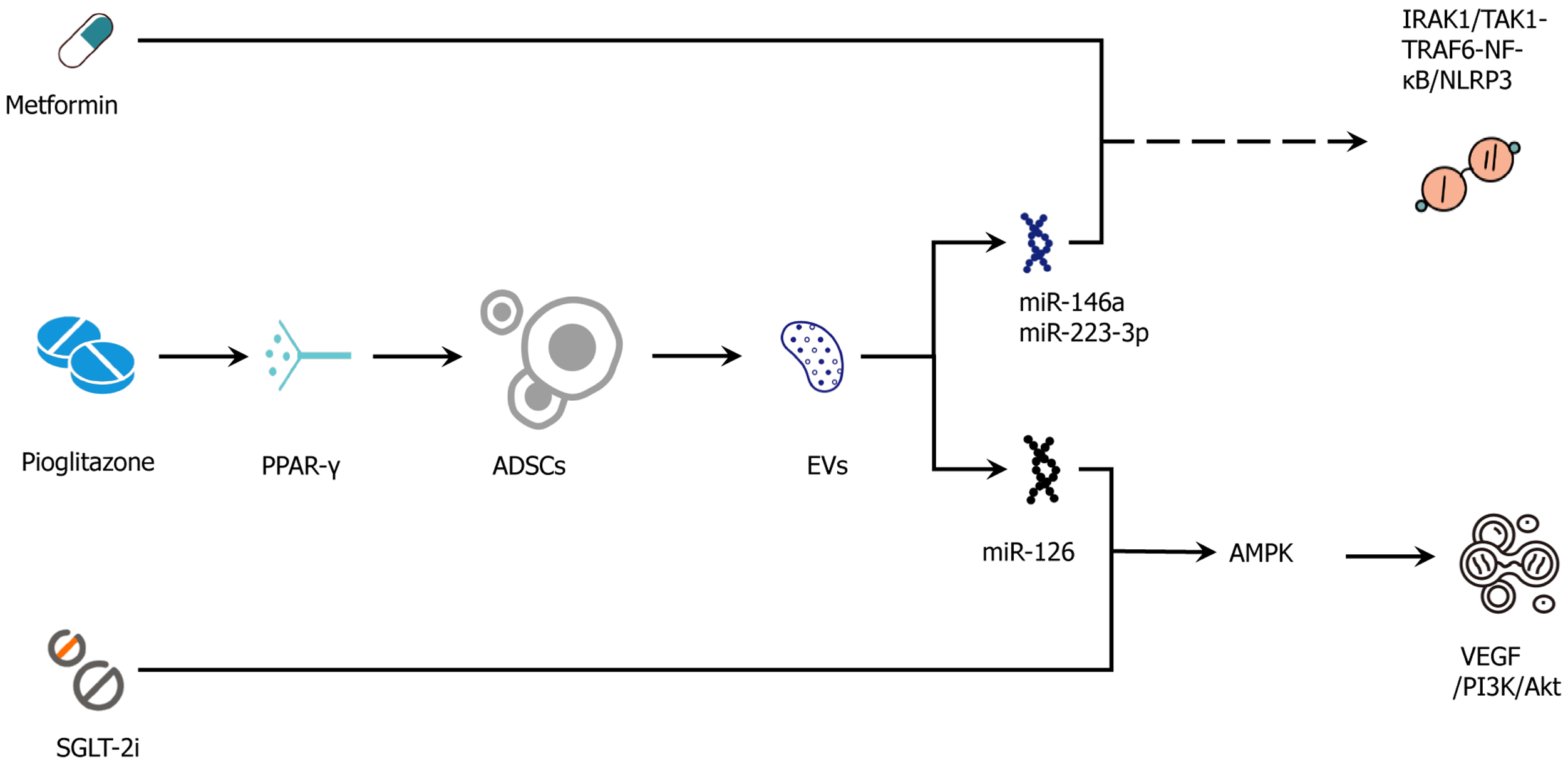
Figure 4 Adipose-derived stem cell-drug synergy map.
Adipose-derived stem cell secretome/extracellular vesicles target drug-action pathways to improve vascular, metabolic, and anti-inflammatory outcomes. Mechanistic intersections whereby adipose-derived stem cell (ADSC) secretome/extracellular vesicles (EVs) augment and complement conventional agents. Metformin (adenosine monophosphate-activated protein kinase) cooperates with ADSC-EV-delivered miR-126 to enhance vascular endothelial growth factor → phosphoinositide 3-kinase/protein kinase B → endothelial nitric oxide synthase signaling and improve endothelial function. Glucagon-like peptide-1 receptor agonist (cyclic adenosine monophosphate/protein kinase A → cyclic adenosine monophosphate response element-binding protein) aligns with ADSC-driven adenosine monophosphate-activated protein kinase/sirtuin-1 → peroxisome proliferator-activated receptor γ coactivator-1α → PR domain zinc finger protein 16/uncoupling protein-1 programs to promote beiging, mitochondrial repair, and fatty-acid oxidation, reducing ceramide/diacylglycerol lipotoxicity. The hemodynamic/anti-inflammatory effects of sodium-glucose cotransporter-2 inhibitors combine with EV miR-146a and miR-223-3p to suppress interleukin-1 receptor-associated kinase-1/transforming growth factor-β-activated kinase-1-tumor necrosis factor receptor-associated factor-6-nuclear factor kappa B → NOD-like receptor protein-3 signaling and reinforce anti-fibrotic responses (mothers against decapentaplegic homolog 2/3 modulation). Peroxisome proliferator-activated receptor γ activation by pioglitazone increases ADSC survival, migration, and vascular endothelial growth factor secretion, providing microenvironmental optimization for subsequent cell/EV therapy. At the insulin-signaling node ADSC effects (suppressor of cytokine signaling-3 ↓ and reduced serine-phosphorylated insulin receptor substrate-1) synergize with drug actions to restore phosphoinositide 3-kinase/protein kinase B/glucose transporter-4 translocation and adipose glucose uptake. These interactions are testable by paired adipose biopsies and serial EV-cargo profiling (e.g., miR-126/146a/223), together with functional readouts (flow-mediated dilation, estimated glomerular filtration rate slope, adipose peroxisome proliferator-activated receptor γ coactivator-1α/uncoupling protein-1, phosphorylated insulin receptor substrate-1). Solid arrows indicate activation/flow; T-bars or dashed lines indicate inhibition or context-dependent routes. IRAK1: Interleukin-1 receptor-associated kinase-1; TAK1: Transforming growth factor-β-activated kinase-1; TRAF6: Tumor necrosis factor receptor-associated factor-6; NF-κB: Nuclear factor kappa B; NLRP3: NOD-like receptor protein-3; PPARγ: Peroxisome proliferator-activated receptor γ; ADSC: Adipose-derived stem cell; EVs: Extracellular vesicles; AMPK: Adenosine monophosphate-activated protein kinase; SGLT-2i: Sodium-glucose cotransporter-2 inhibitor; VEGF: Vascular endothelial growth factor; PI3K: Phosphoinositide 3-kinase; Akt: Protein kinase B; miR: MicroRNA.
- Citation: Luo C, Yu XM, Hua LY, Zeng MQ, Xu H, Duan CZ, Xu SY, Sun D, Ye LY, He DJ. Targeting adipose remodeling: Synergistic mechanisms of drugs and adipose-derived stem cells in obese type 2 diabetes mellitus. World J Stem Cells 2025; 17(11): 111162
- URL: https://www.wjgnet.com/1948-0210/full/v17/i11/111162.htm
- DOI: https://dx.doi.org/10.4252/wjsc.v17.i11.111162