
















©The Author(s) 2025.
World J Stem Cells. Nov 26, 2025; 17(11): 110381
Published online Nov 26, 2025. doi: 10.4252/wjsc.v17.i11.110381
Published online Nov 26, 2025. doi: 10.4252/wjsc.v17.i11.110381
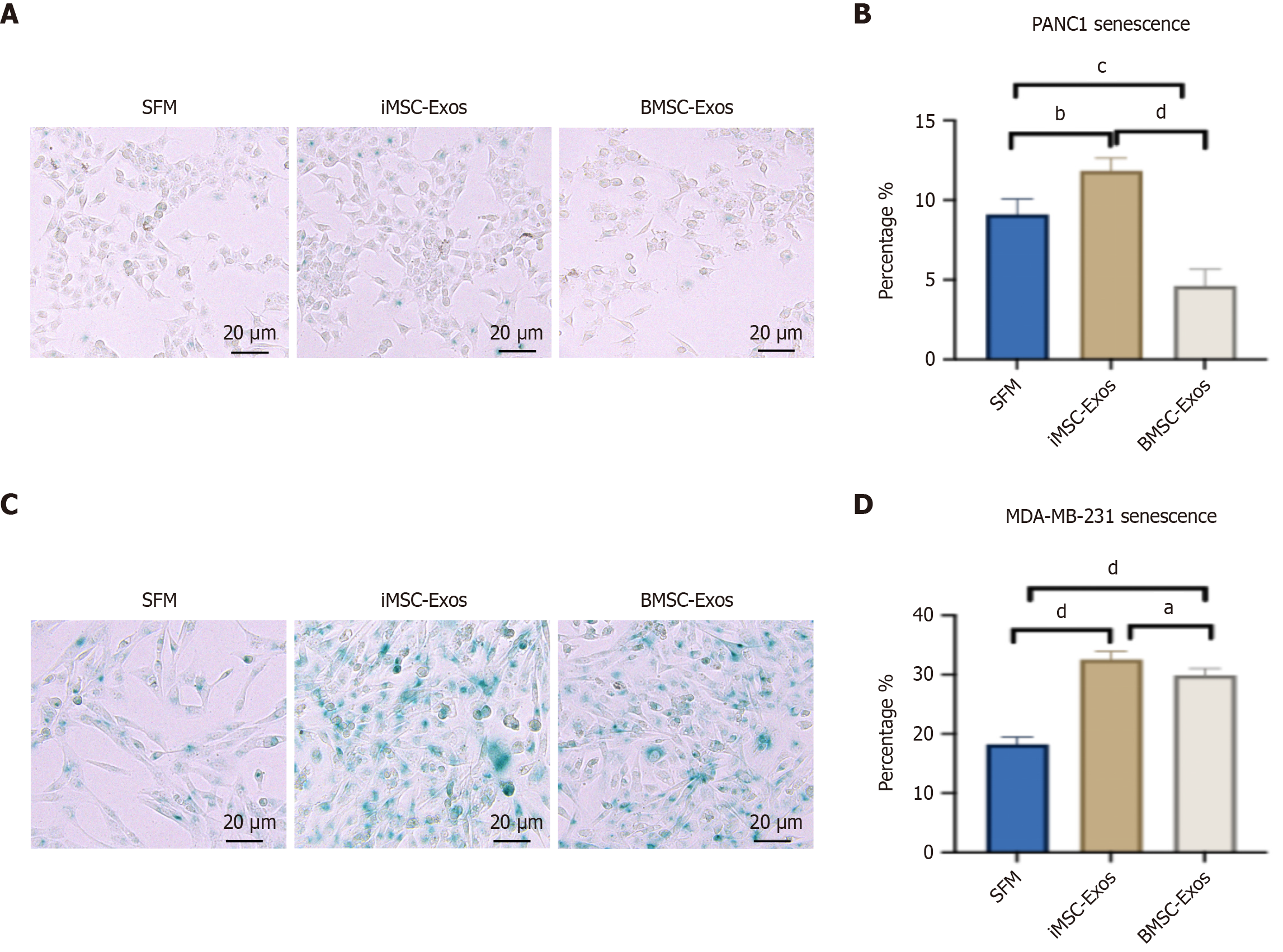
Figure 4 Senescence induction in PANC1 and MDA-MB-231 cells following treatment with exosomes derived from induced pluripotent stem cell-derived mesenchymal stem cells and bone marrow mesenchymal stem cells.
A: Representative images of senescence-associated beta-galactosidase (SA-βGal)-stained PANC1 cells after 48 hours of treatment. Blue staining marks SA-βGal-positive senescent cells (scale bar = 20 μm); B: Quantification of SA-βGal-positive cells expressed as a percentage of the total cell population in PANC1 cultures; C: Representative images of SA-βGal-stained MDA-MB-231 cells after 48 hours of treatment. Blue staining marks SA-βGal-positive senescent cells (scale bar = 20 μm); D: Quantification of SA-βGal-positive cells expressed as a percentage of the total cell population in MDA-MB-231 cultures. Values represent mean ± SD. aP ≤ 0.05, bP ≤ 0.01, cP ≤ 0.001, dP ≤ 0.0001. iMSC-Exos: Exosomes derived from induced pluripotent stem cell-derived mesenchymal stem cells; BMSC-Exos: Exosomes derived from bone marrow mesenchymal stem cell; SFM: Serum-free medium.
- Citation: Ababneh NA, Nashwan S, AlDiqs R, Ismail MA, Abdulelah AA, Abu-Humaidan AHA, AlQirem L, Al-Abdallat K, Al-Qaisi T, Saleh T, Awidi A. Cancer cell-dependent increase in senescence-like populations following exosome treatment from bone marrow and induced pluripotent stem cell-derived mesenchymal stem cells. World J Stem Cells 2025; 17(11): 110381
- URL: https://www.wjgnet.com/1948-0210/full/v17/i11/110381.htm
- DOI: https://dx.doi.org/10.4252/wjsc.v17.i11.110381