
















©The Author(s) 2025.
World J Stem Cells. Nov 26, 2025; 17(11): 110381
Published online Nov 26, 2025. doi: 10.4252/wjsc.v17.i11.110381
Published online Nov 26, 2025. doi: 10.4252/wjsc.v17.i11.110381
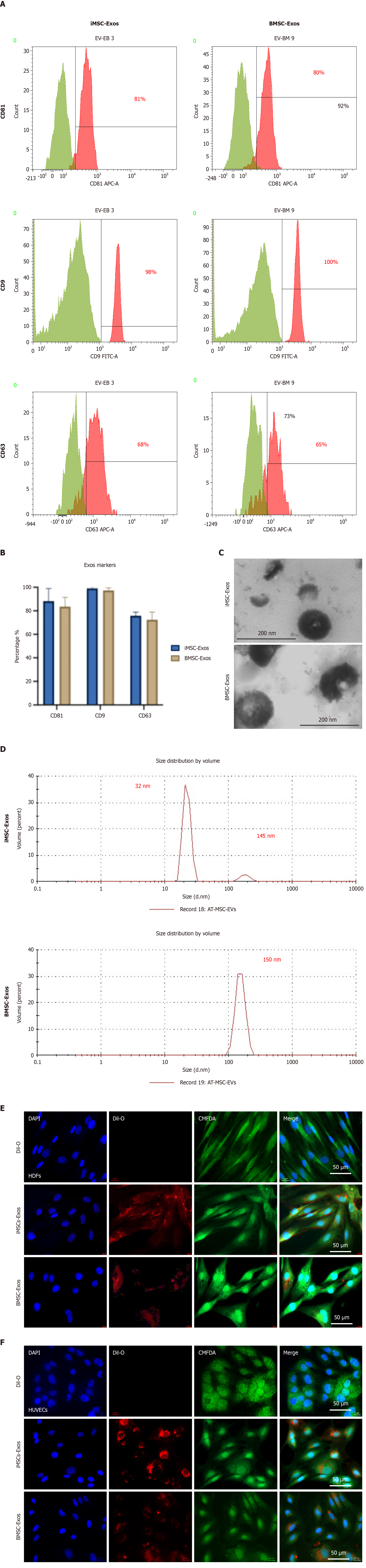
Figure 2 Characterization of exosomes derived from induced pluripotent stem cell-derived mesenchymal stem cells and bone marrow mesenchymal stem cells.
A: Flow cytometry analysis showing expression of tetraspanin surface markers CD9, CD81, and CD63 on exosomes using sulfate-latex bead capture; B: Quantification of surface marker expression comparing exosomes derived from induced pluripotent stem cell-derived mesenchymal stem cells (iMSC-Exos) and exosomes derived from bone marrow mesenchymal stem cells (BMSC-Exos); C: Representative transmission electron microscopy images of iMSC-Exos (left) and BMSC-Exos (right). Scale bar = 200 nm; D: Dynamic light scattering profiles depicting size distribution of iMSC-Exos (top) and BMSC-Exos (bottom); E and F: Confocal microscopy images showing uptake of DiI-labeled exosomes (red) by human dermal fibroblasts (E) and human umbilical vein endothelial cells (F). Cells were costained with 5-chloromethylfluorescein diacetate (green) for the cytoplasm and DAPI (blue) for the nuclei. Scale bar = 50 μm. iMSC-Exos: Exosomes derived from induced pluripotent stem cell-derived mesenchymal stem cells; BMSC-Exos: Exosomes derived from bone marrow mesenchymal stem cell; Exos: Exosomes; HDFs: Human dermal fibroblasts; HUVECs: Human umbilical vein endothelial cells.
- Citation: Ababneh NA, Nashwan S, AlDiqs R, Ismail MA, Abdulelah AA, Abu-Humaidan AHA, AlQirem L, Al-Abdallat K, Al-Qaisi T, Saleh T, Awidi A. Cancer cell-dependent increase in senescence-like populations following exosome treatment from bone marrow and induced pluripotent stem cell-derived mesenchymal stem cells. World J Stem Cells 2025; 17(11): 110381
- URL: https://www.wjgnet.com/1948-0210/full/v17/i11/110381.htm
- DOI: https://dx.doi.org/10.4252/wjsc.v17.i11.110381