
















©The Author(s) 2025.
World J Stem Cells. Oct 26, 2025; 17(10): 111792
Published online Oct 26, 2025. doi: 10.4252/wjsc.v17.i10.111792
Published online Oct 26, 2025. doi: 10.4252/wjsc.v17.i10.111792
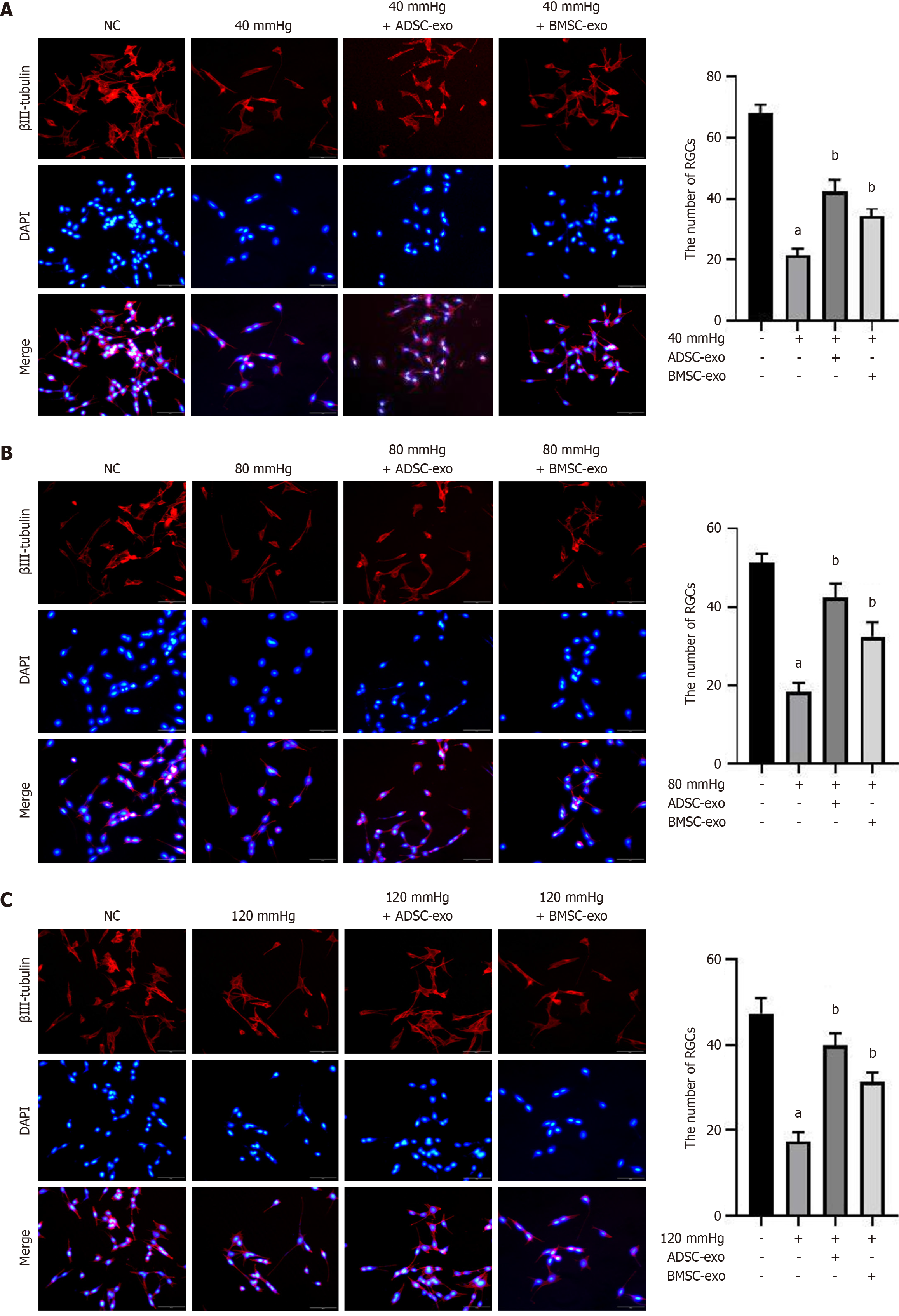
Figure 4 Immunofluorescence detection of p-CREB and CREB.
A-C: Immunofluorescence staining of CREB (green) and p-CREB (red) and nuclear staining with DAPI (blue) (left panel) and quantification of the p-CREB/CREB ratio (right panel) in different groups after exposure to (A) 40 mmHg, (B) 80 mmHg, and (C) 120 mmHg (scale bar: 20 μm). aP < 0.05, compared with the normal control group; bP < 0.05, compared with the model (40, 80, and 120 mmHg) group. ADSC: Adipose-derived stem cell; BMSC: Bone marrow-derived stem cell; Exo: Exosome; NC: Normal control.
- Citation: Dai M. Correction to “ADSC-Exos outperform BMSC-Exos in alleviating hydrostatic pressure-induced injury to retinal ganglion cells by upregulating nerve growth factors”. World J Stem Cells 2025; 17(10): 111792
- URL: https://www.wjgnet.com/1948-0210/full/v17/i10/111792.htm
- DOI: https://dx.doi.org/10.4252/wjsc.v17.i10.111792