
















©The Author(s) 2025.
World J Stem Cells. Oct 26, 2025; 17(10): 110445
Published online Oct 26, 2025. doi: 10.4252/wjsc.v17.i10.110445
Published online Oct 26, 2025. doi: 10.4252/wjsc.v17.i10.110445
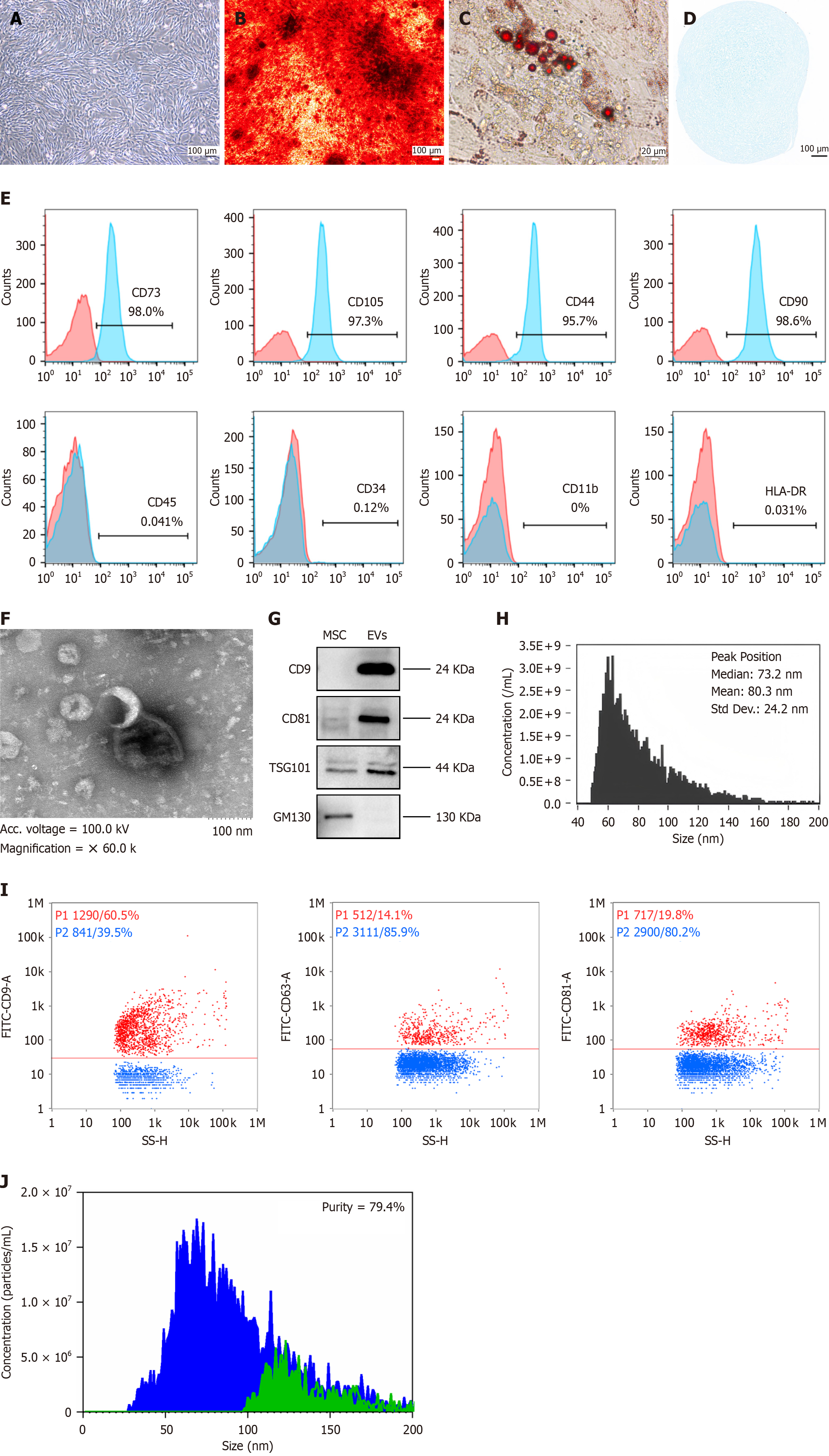
Figure 1 The characteristics of umbilical cord-derived mesenchymal stromal cells and derived extracellular vesicles derived from mesenchymal stromal cells.
A: Morphological examination of umbilical cord-derived mesenchymal stromal cells (UC-MSCs) (scale bar indicates 100 μm); B-D: Multilineage differentiation of UC-MSCs. B: Osteogenic differentiation characterized by calcium phosphate deposits, visualized by Alizarin Red staining (scale bar indicates 100 μm); C: Adipogenic differentiation confirmed by the accumulation of lipid vacuoles, highlighted by Oil Red O staining (scale bar indicates 20 μm); D: Chondrogenic differentiation indicated by the synthesis of proteoglycans stained with Alcian Blue (scale bar indicates 100 μm); E: Surface marker expression profile of UC-MSCs. These cells were positive for the mesenchymal markers CD73 (98.0%), CD105 (97.3%), CD44 (95.7%), and CD90 (98.6%) but negative for CD45 (0.041%), CD34 (0.12%), CD11b (0.0%), and HLA-DR (0.031%); F: The ultrastructure of extracellular vesicles (EVs) derived from mesenchymal stromal cells (MSC-EVs) was examined by transmission electron microscopy (the scale bar indicates 100 nm); G: Immunoblotting was performed for the biomarkers of UC-MSCs and MSC-EVs. Full-length blots are presented in Supplementary Figure 4; H: The histogram illustrates the particle size distribution of MSC-EVs. To evaluate the concentration and dimensional spread of the particles, the suspension containing EVs was diluted at a 1:1000 ratio with phosphate buffered saline; I: The CD9, CD63, and CD81 proteins expressed on the surface of MSC-EVs were detected by nano flow cytometry. For immunofluorescence staining, fluorescein isothiocyanate-labeled antibodies against CD9, CD63, or CD81 were added to the EV suspension. All antibodies were purchased from BD Biosciences; J: Comparative particle quantification of MSC-EVs was performed before and after treatment with Triton X-100. To assess purity, a 5 μL aliquot of 10% Triton X-100 (purchased from Sigma-Aldrich, Japan) was mixed with a 45 μL suspension of EVs. The nano flow cytometry platform was used to analyze the characteristic side scatter burst profiles of the EVs. Purity = (pre-disruption particle count - post-disruption particle count)/pre-disruption particle count × 100%, higher values indicate greater purity. MSCs: Mesenchymal stromal cells; EVs: Extracellular vesicles; TSG101: Tumor susceptibility gene 101.
- Citation: Yang SS, Chen SY, Zhuang WY, Han J, Liu Y, Deng L, Guo HZ, Ma HR, Tan Y. Efficacy of extracellular vesicles derived from mesenchymal stromal cells in regulating senescence: In vitro and in vivo insights. World J Stem Cells 2025; 17(10): 110445
- URL: https://www.wjgnet.com/1948-0210/full/v17/i10/110445.htm
- DOI: https://dx.doi.org/10.4252/wjsc.v17.i10.110445