
















©The Author(s) 2025.
World J Stem Cells. Oct 26, 2025; 17(10): 110190
Published online Oct 26, 2025. doi: 10.4252/wjsc.v17.i10.110190
Published online Oct 26, 2025. doi: 10.4252/wjsc.v17.i10.110190
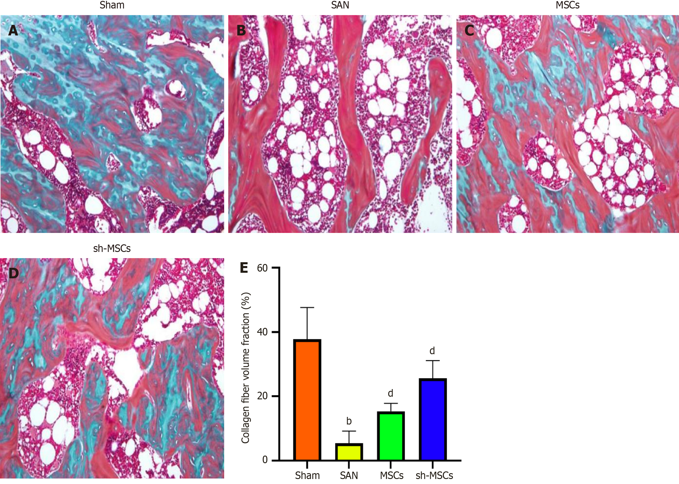
Figure 5 Sclerostin inhibitor-silenced human umbilical cord mesenchymal stem cells promote new bone formation in mice with steroid-induced avascular necrosis of the femoral head.
A and B: Masson staining revealed distinct bone repair patterns: Sham group showed prominent blue-stained new bone areas, indicating active repair (A), SAN group exhibited predominantly red-stained old bone with minimal blue areas, suggesting impaired repair (B); C and D: Both MSCs-treated groups displayed osteoblasts, with sh-MSCs group (D) showing more extensive blue staining than MSCs group (C); E: Quantification demonstrated significantly reduced collagen fiber volume in SAN group vs sham (P < 0.01). MSC treatments restored collagen content, with sh-MSCs showing superior improvement vs MSCs (P < 0.01 vs SAN). Data represent mean ± SEM from three independent experiments. bP < 0.01 vs sham; dP < 0.01 vs SAN. SAN: Steroid-induced avascular necrosis of the femoral head; MSC: Mesenchymal stem cell.
- Citation: Lv H, Zheng CF, Chen XY, Wei JH, Tao YZ, Feng L, Feng Z, Lu SJ. Sclerostin-silenced human umbilical cord mesenchymal stem cells ameliorate bone metabolism in steroid-induced femoral head necrosis. World J Stem Cells 2025; 17(10): 110190
- URL: https://www.wjgnet.com/1948-0210/full/v17/i10/110190.htm
- DOI: https://dx.doi.org/10.4252/wjsc.v17.i10.110190