
















©The Author(s) 2025.
World J Stem Cells. Oct 26, 2025; 17(10): 110190
Published online Oct 26, 2025. doi: 10.4252/wjsc.v17.i10.110190
Published online Oct 26, 2025. doi: 10.4252/wjsc.v17.i10.110190
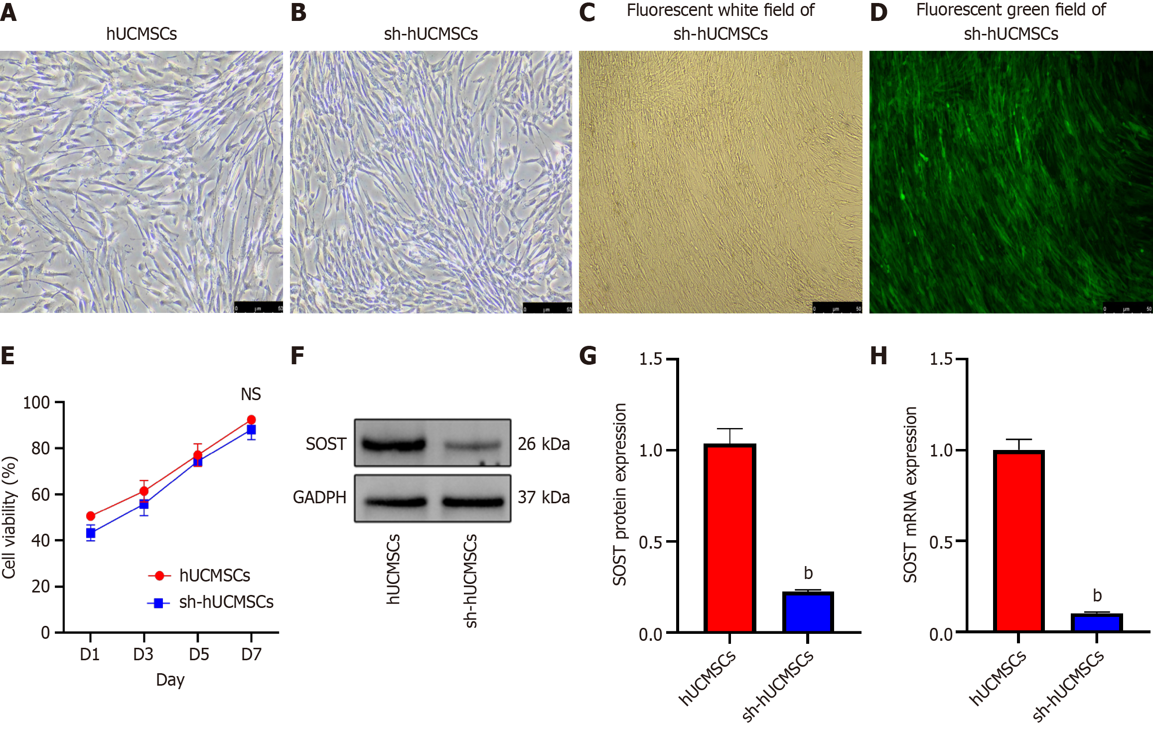
Figure 2 Human umbilical cord mesenchymal stem cells and their morphology after electroporation.
Morphology and transfection efficiency of human umbilical cord mesenchymal stem cells. A: Passage 3 (P3) human umbilical cord mesenchymal stem cells (hUCMSCs) showing spindle-shaped, radially arranged growth (scale bar = 50 μm); B: P3 hUCMSCs post-electroporation with sclerostin (SOST) inhibitor plasmid (scale bar = 50 μm); C and D: P4 SOST-RNAi hUCMSCs showing stable green fluorescent protein expression after puromycin selection (C: Bright field; D: Fluorescence field), scale bar = 50 μm; E: The cell viability of each group of hUCMSCs; F: Western blot of SOST gene silencing; G: SOST protein expression levels were significantly decreased in hUCMSCs with SOST-silenced; H: SOST mRNA expression levels were significantly decreased in hUCMSCs with SOST-silenced via quantitative real-time polymerase chain reaction. All data were presents as mean ± SD, n = 3. bP < 0.01 vs human umbilical cord mesenchymal stem cells; NS: Not significant. SOST: Sclerostin; hUCMSC: Human umbilical cord mesenchymal stem cells; sh-hUCMSCs: Human umbilical cord mesenchymal stem cells with sclerostin-silenced.
- Citation: Lv H, Zheng CF, Chen XY, Wei JH, Tao YZ, Feng L, Feng Z, Lu SJ. Sclerostin-silenced human umbilical cord mesenchymal stem cells ameliorate bone metabolism in steroid-induced femoral head necrosis. World J Stem Cells 2025; 17(10): 110190
- URL: https://www.wjgnet.com/1948-0210/full/v17/i10/110190.htm
- DOI: https://dx.doi.org/10.4252/wjsc.v17.i10.110190