
















©The Author(s) 2025.
World J Stem Cells. Oct 26, 2025; 17(10): 109862
Published online Oct 26, 2025. doi: 10.4252/wjsc.v17.i10.109862
Published online Oct 26, 2025. doi: 10.4252/wjsc.v17.i10.109862
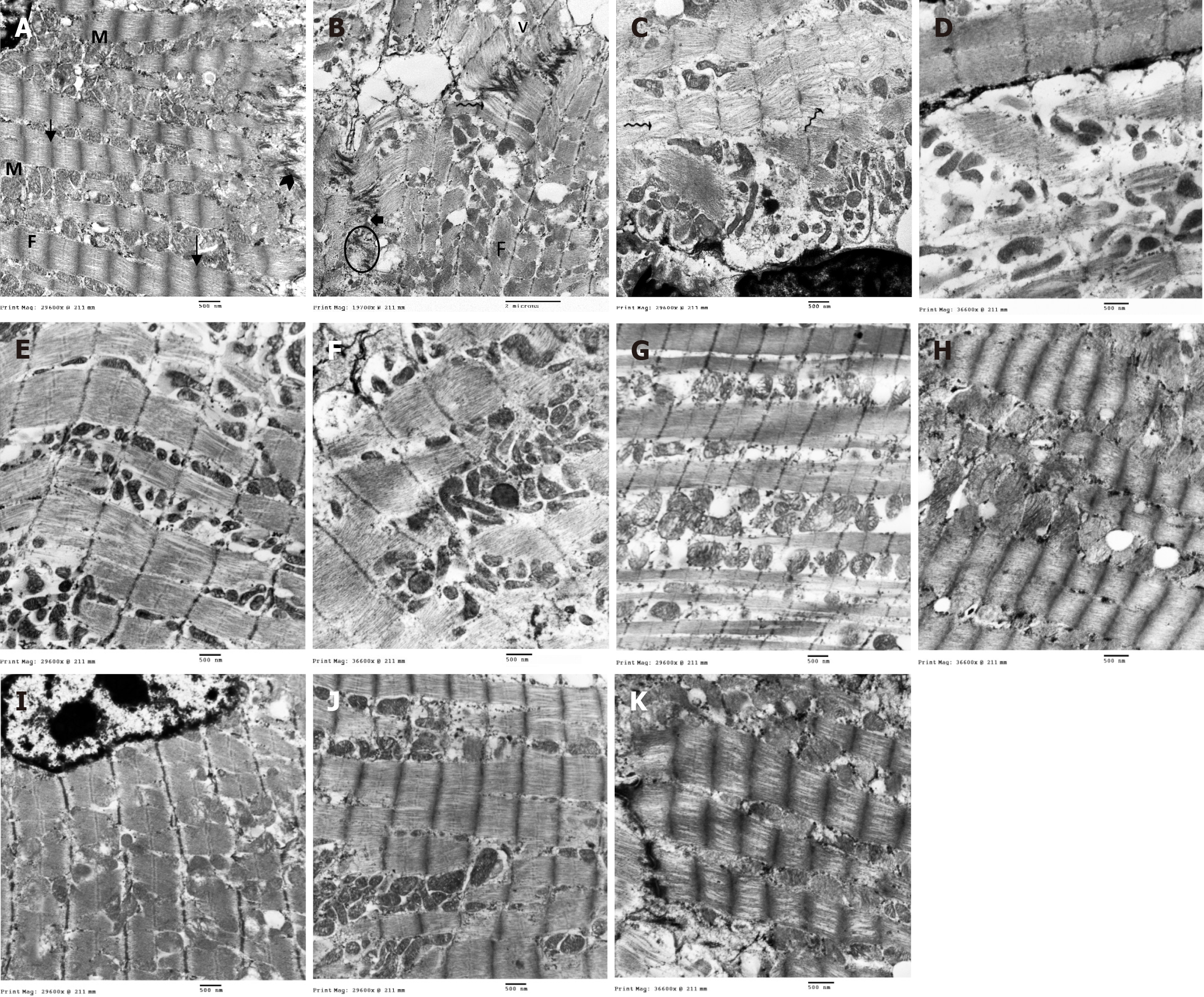
Figure 9 Ultrastructure of cardiac muscle fibers in different groups.
A: Control group: Normal mitochondrial (M) distribution in linear arrays alongside the myofibrils. Mitochondria are electron dense with tightly packed tubular cristae. Myofibrils (F) being formed of regular sarcomeres separated by Z lines (arrow) and attached to the transverse portion of the intercalated disc (arrowhead); B-D: ISO + MI group: Markedly irregular myofibrils (F) with separation of myofilaments and in-between the myofilaments (wavy arrow), numerous irregular and bizarre-shaped mitochondria (M) with extensive disruption of mitochondrial cristae, large vacuolations (v), widening of sarcoplasmic reticulum tubules (curved arrow), desmosome paleness and widening of adherens junction gaps (short arrow), clumping of nuclear chromatin (N), widening of sub-sarcolemma; E and F: MI + EXE group: Closely packed myofibrils with regular sarcomeres and mitochondria arranged in-between; some of them with disrupted cristae others with irregular shapes, sub-sarcolemmal widening, and areas of focal loss of some myofilaments; G and H: MI + MSC-EXO group: Closely packed myofibrils with regular sarcomeres, yet swollen mitochondria (M), dilated sarcoplasmic reticulum cisternae (curved arrow) and some glycogen granules are noted; I-K: MI + EXE + MSC-EXO group: Closely packed myofibrils with regular sarcomeres and attached to the transverse portion of the intercalated disc, numerous mitochondria filled with intact cristae and abundant glycogen granules (magnifications A, C, E, G and I: 12000 ×; B: 8000 ×; D, F, H and J: 15000 ×).
- Citation: ShamsEldeen AM, AbdElalim MM, Mohamed NS, AbdelRahman MM, Kamar SS, AbdelKader DH, Elsharkawy SH, Rashed LA, Faisal SS, Osman WA, Selmy AM. Mesenchymal stem-derived exosomes enhance therapeutic benefits of exercise in isoproterenol-induced myocardial ischemia: Targeting ERK and Akt/mTOR signaling. World J Stem Cells 2025; 17(10): 109862
- URL: https://www.wjgnet.com/1948-0210/full/v17/i10/109862.htm
- DOI: https://dx.doi.org/10.4252/wjsc.v17.i10.109862