
















©The Author(s) 2025.
World J Stem Cells. Oct 26, 2025; 17(10): 109862
Published online Oct 26, 2025. doi: 10.4252/wjsc.v17.i10.109862
Published online Oct 26, 2025. doi: 10.4252/wjsc.v17.i10.109862
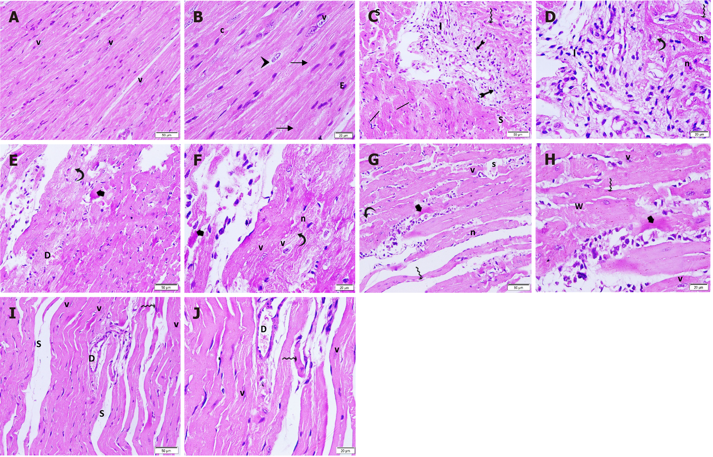
Figure 7 Hematoxylin and eosin-stained longitudinal sections in the cardiac tissue in different groups.
A and B: Control group: Branching muscle fibers with striated acidophilic cytoplasm. The delicate connective tissue in between the muscle fibers contain blood capillaries; C and D: ISO + MI group: Large focal intramural pale stained-area infiltrated with mononuclear cells and fibroblast illustrating spindle-shaped flattened nuclei. The surrounding muscle fibers show increased diameter; E and F: MI + EXE group: A small focal area of fragmented muscle fibers with few intercellular mononuclear cell infiltrations. Surrounding viable muscle fibers are noted while others display a wavy appearance; G and H: MI + MSC-EXO group: A small focal area of degenerated muscle fibers with few intercellular mononuclear cell infiltrations is noted surrounded by some viable muscle fibers; I and J: MI + EXE + MSC-EXO group: Few thinned-out muscle fibers with separation in-between are noted surrounded by many viable closely and regularly adjusted cardio-myocytes (magnifications A, C, E, G and I: 200 ×; B, D, F, H and J: 400 ×). v: Vesicular nuclei; arrowhead: Perinuclear pale area; arrow: Intercalated discs; c: Blood capillaries; E: Endothelium; I: Inflammatory infiltration; bifid arrow: Fibroblast; wavy arrow: Thinned-out muscle fibers; line segment: Dilated muscle fibers; curved arrow: Vacuolated cytoplasm; n: Dark shrunken nuclei; S: Separation; short arrow: Dense acidophilic cytoplasm; w: Wavy muscle fibers; D: Dilated blood vessel.
- Citation: ShamsEldeen AM, AbdElalim MM, Mohamed NS, AbdelRahman MM, Kamar SS, AbdelKader DH, Elsharkawy SH, Rashed LA, Faisal SS, Osman WA, Selmy AM. Mesenchymal stem-derived exosomes enhance therapeutic benefits of exercise in isoproterenol-induced myocardial ischemia: Targeting ERK and Akt/mTOR signaling. World J Stem Cells 2025; 17(10): 109862
- URL: https://www.wjgnet.com/1948-0210/full/v17/i10/109862.htm
- DOI: https://dx.doi.org/10.4252/wjsc.v17.i10.109862