
















©The Author(s) 2025.
World J Stem Cells. Oct 26, 2025; 17(10): 109862
Published online Oct 26, 2025. doi: 10.4252/wjsc.v17.i10.109862
Published online Oct 26, 2025. doi: 10.4252/wjsc.v17.i10.109862
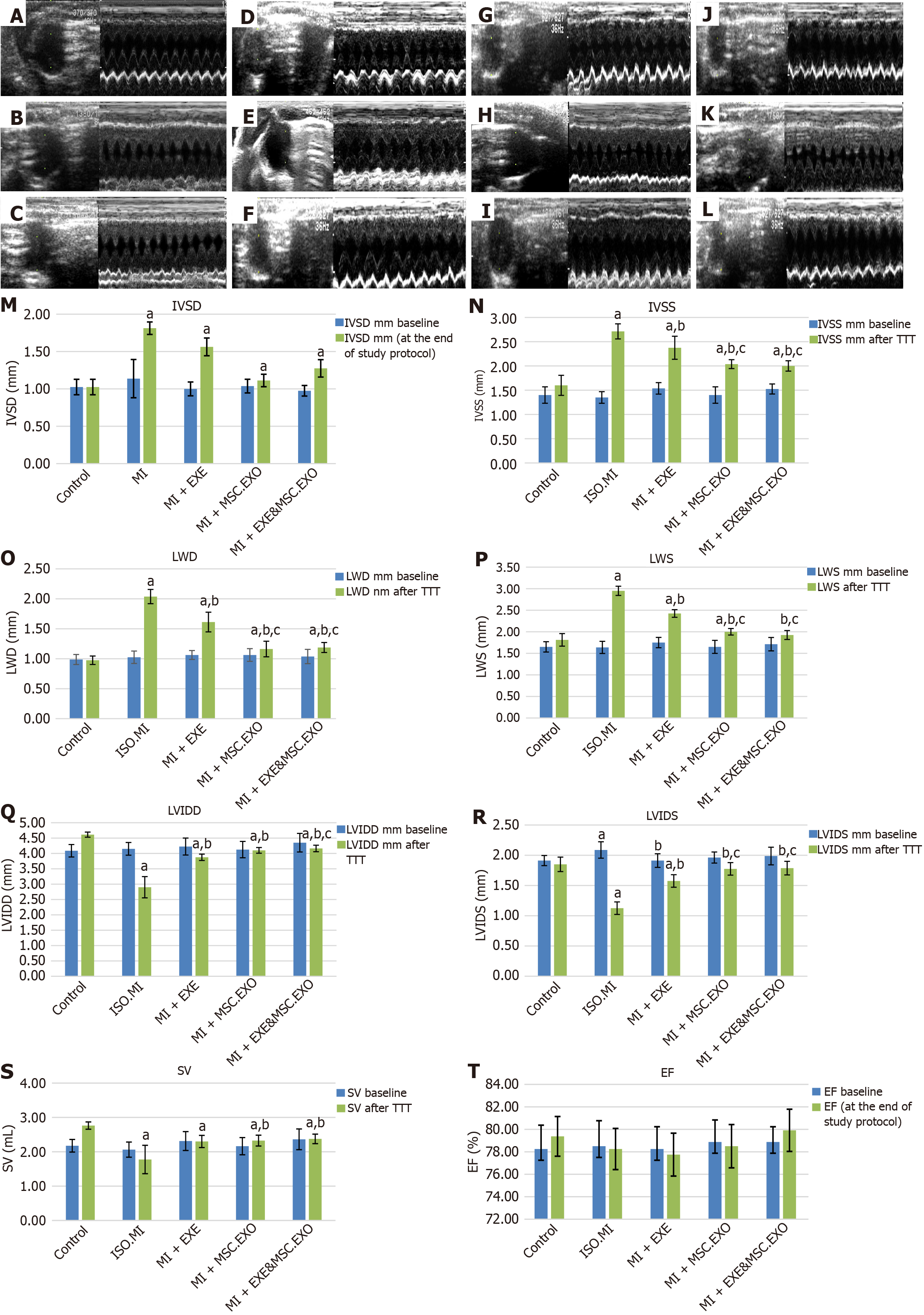
Figure 4 Comparison of different estimated ECHO parameters and quantification of the estimated ECHO parameters.
A-L: Echocardiographic examination guided by right parasternal short-axis view at the level of the papillary muscle (M-mode). ISO + MI group (A-C): At the start, it shows normal septal and free wall thickness in control rats (A), ISO + MI group shows marked septal and free wall hypertrophy (B), after the end of the study protocol, preserved septal and free wall hypertrophy is noticed (C); MI + EXE group (D-F): Showing normal wall thickness (D), marked septal and free wall hypertrophy in group MI + EXE just after induction of MI by ISO (E), septal and free wall hypertrophy subsided after exercise training for 4 weeks (F); MI + MSC-EXO group (G-I): Normal septal and free wall thickness in control rats (G), moderate septal and free wall hypertrophy is noticed in MI + MSC-EXO after induction of MI (H), full regression of the wall is observed after receiving MSC-EXO (I); MI + EXE + MSC-EXO group (J-L): Normal wall thickness in the control group (J), septal and free wall hypertrophy is noticed in MI + EXE + MSC-EXO rats after induction of MI (K), fully regressed after receiving both EXE and MSC-EXO regimen (L); M-T: Quantification of the estimated ECHO parameters: Interventricular septal diameter during diastole (M), interventricular septal diameter during systole (N), lateral wall diameter during diastole (O), lateral wall diameter during systole (P), left ventricular internal diameter during diastole (Q), left ventricular internal diameter during systole (R), stroke volume (S), and ejection fraction% (T). Values represent means ± SD (n = 8). aP < 0.05, compared to the control group, bP < 0.05, compared to ISO + MI group, cP < 0.05, compared to MI + EXE group, dP < 0.05, compared to MI + MSC-EXO group. IVSD: Interventricular septal diameter during diastole; IVSS: Interventricular septal diameter during systole; LWD: Lateral wall diameter during diastole; LWS: Lateral wall diameter during systole; LVIDD: Left ventricular internal diameter during diastole; LVIDS: Left ventricular internal diameter during systole; SV: Stroke volume; EF: Ejection fraction; MSC-EXO: Mesenchymal stem cell-derived exosomes.
- Citation: ShamsEldeen AM, AbdElalim MM, Mohamed NS, AbdelRahman MM, Kamar SS, AbdelKader DH, Elsharkawy SH, Rashed LA, Faisal SS, Osman WA, Selmy AM. Mesenchymal stem-derived exosomes enhance therapeutic benefits of exercise in isoproterenol-induced myocardial ischemia: Targeting ERK and Akt/mTOR signaling. World J Stem Cells 2025; 17(10): 109862
- URL: https://www.wjgnet.com/1948-0210/full/v17/i10/109862.htm
- DOI: https://dx.doi.org/10.4252/wjsc.v17.i10.109862